Japanese
English
- 有料閲覧
- Abstract 文献概要
- 1ページ目 Look Inside
I.はじめに
脳神経外科における顕微鏡下手術では,術者は術野を直接見るのではなく,術野の拡大映像が呈示される.また,手術操作は器具を通して遠隔的に行われる.これは一種のバーチャルリアリティー(VR)環境とも考えうるが,外科医の感覚と操作間のギャップにより,操作の動揺や術野方向感覚の喪失がもたらされる.これまで,このようなギャップを克服することが臨床修練の一環とみなされてきた.今後,ナビゲータ下手術などVR化が進むにつれ1,2,10)ギャップは質的に変化しつつ拡大すると考えられる.本研究では私たちが開発した脳手術ナビゲータ「CANS Navigator」3,4,6)を用いて種々のVR環境下で術野ファントムにおける到達過程を記録,比較分析し,術者の空間認識と手術操作間のギャップの解明を試み,ついでこのギャップを改善しうる適切なインターフェイスの考案を試みた.
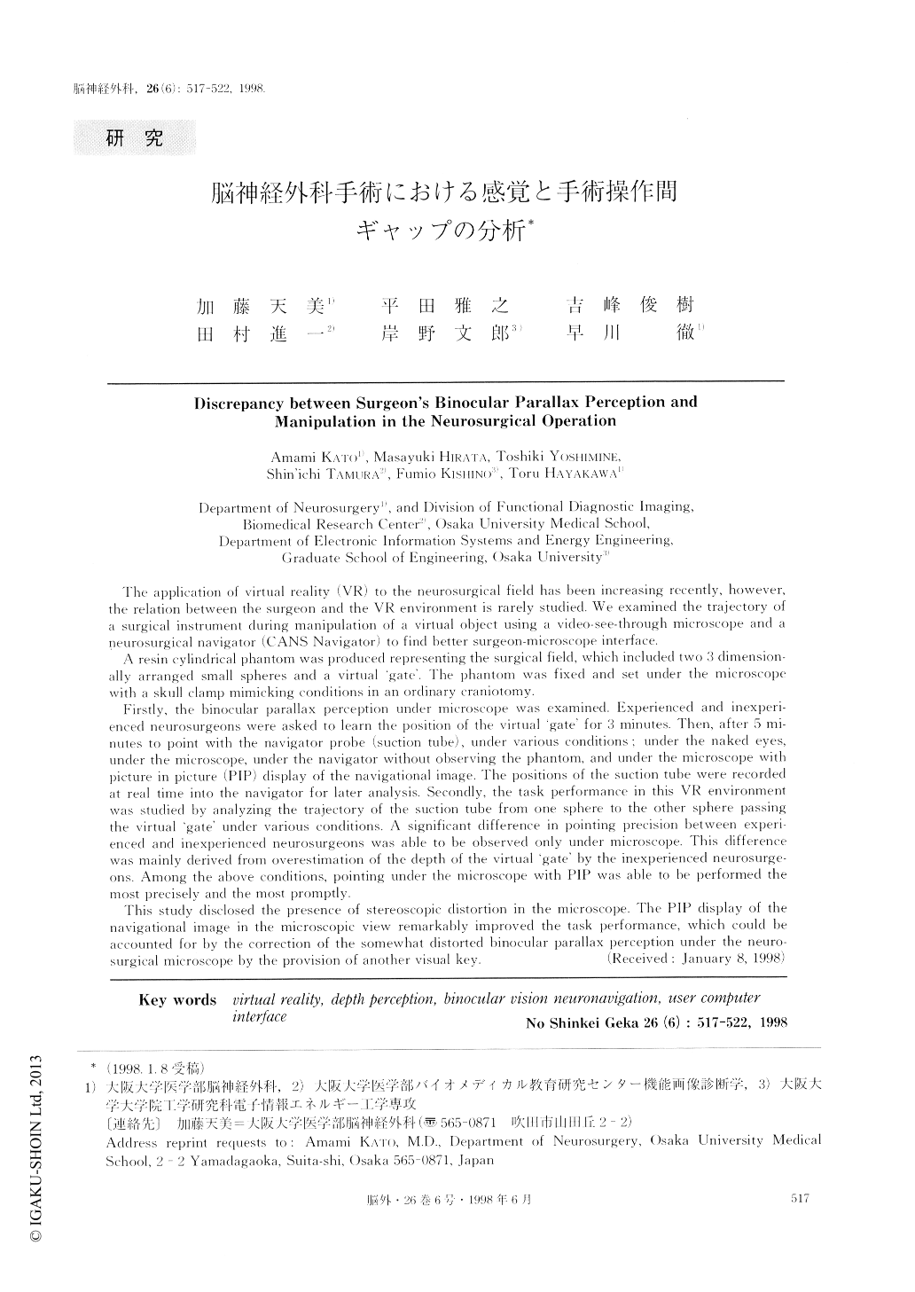
The application of virtual reality (VR) to the neurosurgical field has been increasing recently, however,the relation between the surgeon and the VR environment is rarely studied. We examined the trajectory ofa surgical instrument during manipulation of a virtual object using a video-see-through microscope and aneurosurgical navigator (CANS Navigator) to find better surgeon-microscope interface.
A resin cylindrical phantom was produced representing the surgical field, which included two 3 dimension-ally arranged small spheres and a virtual 'gate'. The phantom was fixed and set under the microscopewith a skull clamp mimicking conditions in an ordinary craniotomy.
Firstly, the binocular parallax perception under microscope was examined. Experienced and inexperi-enced neurosurgeons were asked to learn the position of the virtual 'gate' for 3 minutes. Then, after 5 mi-nutes to point with the navigator probe (suction tube), under various conditions; under the naked eyes,under the microscope, under the navigator without observing the phantom, and under the microscope withpicture in picture (PIP) display of the navigational image. The positions of the suction tube were recordedat real time into the navigator for later analysis. Secondly, the task performance in this VR environmentwas studied by analyzing the trajectory of the suction tube from one sphere to the other sphere passingthe virtual 'gate' under various conditions. A significant difference in pointing precision between experi-enced and inexperienced neurosurgeons was able to he observed only under microscope. This differencewas mainly derived from overestimation of the depth of the virtual 'gate' by the inexperienced neurosurge-ons. Among the above conditions, pointing under the microscope, with PIP was able to be performed themost precisely and the most promptly.
This study disclosed the presence of stereoscopic distortion in the microscope. The PIP display of thenavigational image in the microscopic view remarkably improved the task performance, which could beaccounted for by the correction of the somewhat distorted binocular parallax perception under the neuro-surgical microscope by the provision of another visual key.

Copyright © 1998, Igaku-Shoin Ltd. All rights reserved.


