Japanese
English
症例
経皮的生検により診断した腎盂白板症
A CASE OF LEUKOPLAKIA OF RENAL PELVIS DIAGNOSED BY PERCUTANEOUS TECHNOLOGY
仲田 浄治郎
1
,
増田 富士男
1
,
飯塚 典男
1
,
近藤 泉
1
,
五十嵐 宏
1
,
町田 豊平
1
Jojiro Nakada
1
,
Fujio Masuda
1
,
Norio Iizuka
1
,
Izumi Kondo
1
,
Hiroshi Igarashi
1
,
Toyohey Machida
1
1東京慈恵会医科大学泌尿器科学教室
1Depertment of Urology, The Jikei University School of Medicine
キーワード:
腎盂白板症
Keyword:
腎盂白板症
pp.335-337
発行日 1988年4月20日
Published Date 1988/4/20
DOI https://doi.org/10.11477/mf.1413204722
- 有料閲覧
- Abstract 文献概要
- 1ページ目 Look Inside
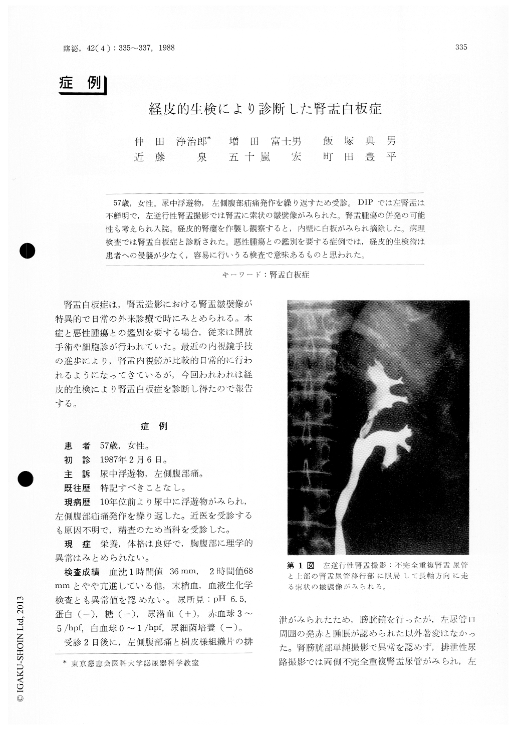
57歳,女性。尿中浮遊物,左側腹部疝痛発作を繰り返すため受診。DIPでは左腎盂は不鮮明で,左逆行性腎盂撮影では腎盂に索状の皺襞像がみられた。腎盂腫瘍の併発の可能性も考えられ入院。経皮的腎瘻を作製し観察すると,内壁に白板がみられ摘除した。病理検査では腎盂白板症と診断された。悪性腫瘍との鑑別を要する症例では,経皮的生検術は患者への侵襲が少なく,容易に行いうる検査で意味あるものと思われた。
A 57-year-old women visited our department because of cloudy urine and repeated left renal colic pain. DIP revealed obscure left renal pelvis. Left retrograde pyelography showed stringy filling defects in the renal pelvis. Because of possible complication with renal pelvic tumor percutaneous nephrostomy was established and observation was made. Leukoplakia was found on the renal pelvic wall, which was resected. Pathological diagnosis was leukoplakia of the renal pelvis.

Copyright © 1988, Igaku-Shoin Ltd. All rights reserved.


