Japanese
English
原著
骨盤腔および後腹膜腔におけるdirect coronal CTの試み
DIRECT CORONAL COMPUTED TOMOGRAPHY OF THE PELVIS AND THE RETROPERITONEAL SPACE
梅山 知一
1
,
根本 良介
1
,
根本 真一
1
,
石川 悟
1
,
矢崎 恒忠
1
,
林正 健二
1
,
加納 勝利
1
,
北川 龍一
1
,
殿岡 怜子
2
,
平松 慶博
2
Tomokazu Umeyama
1
,
Ryosuke Nemoto
1
,
Shinichi Nemoto
1
,
Satoru Ishikawa
1
,
Tsunetada Yazaki
1
,
Kenji Rinsho
1
,
Shori Kanoh
1
,
Ryuichi Kitagawa
1
,
Reiko Tonooka
2
,
Yoshihiro Hiramatsu
2
1筑波大学臨床医学系泌尿器科
2筑波大学臨床医学系放射線科
1Department of Urology, Institute of Clinical Medicine, University of Tsukuba
2Department of Radiology, Institute of Clinical Medicine, University of Tsukuba
pp.427-431
発行日 1983年5月20日
Published Date 1983/5/20
DOI https://doi.org/10.11477/mf.1413203572
- 有料閲覧
- Abstract 文献概要
- 1ページ目 Look Inside
緒言
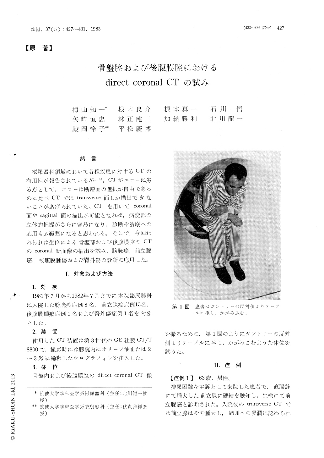
泌尿器科領域において各種疾患に対するCTの有用性が報告されているが1〜4),CTがエコーに劣る点として,エコーは断層面の選択が自由であるのに比べCTではtransverse面しか描出できないことがあげられていた。CTを用いてcoronal面やsagittal面の描出が可能となれば,病変部の立体的把握がさらに容易になり,診断や治療への応用も広範囲になると思われる。そこで,今回われわれは坐位による骨盤部および後腹膜腔のCTのcoronal断面像の描出を試み,膀胱癌,前立腺癌,後腹膜腫瘍および腎外傷の診断に応用した。
In comparison with ultrasonography, the disadvantage of the technique of computed tomography (CT) has been the relative lack of flexibility afforded by the single transverse plane of section. Therefore we developed direct coronal CT imaging of the pelvis and the retroperitoneal space by changing the position of the patient in the gantry. In comparison with transverse CT images we got some advantages in this method.

Copyright © 1983, Igaku-Shoin Ltd. All rights reserved.


