Japanese
English
- 有料閲覧
- Abstract 文献概要
- 1ページ目 Look Inside
- 参考文献 Reference
はじめに
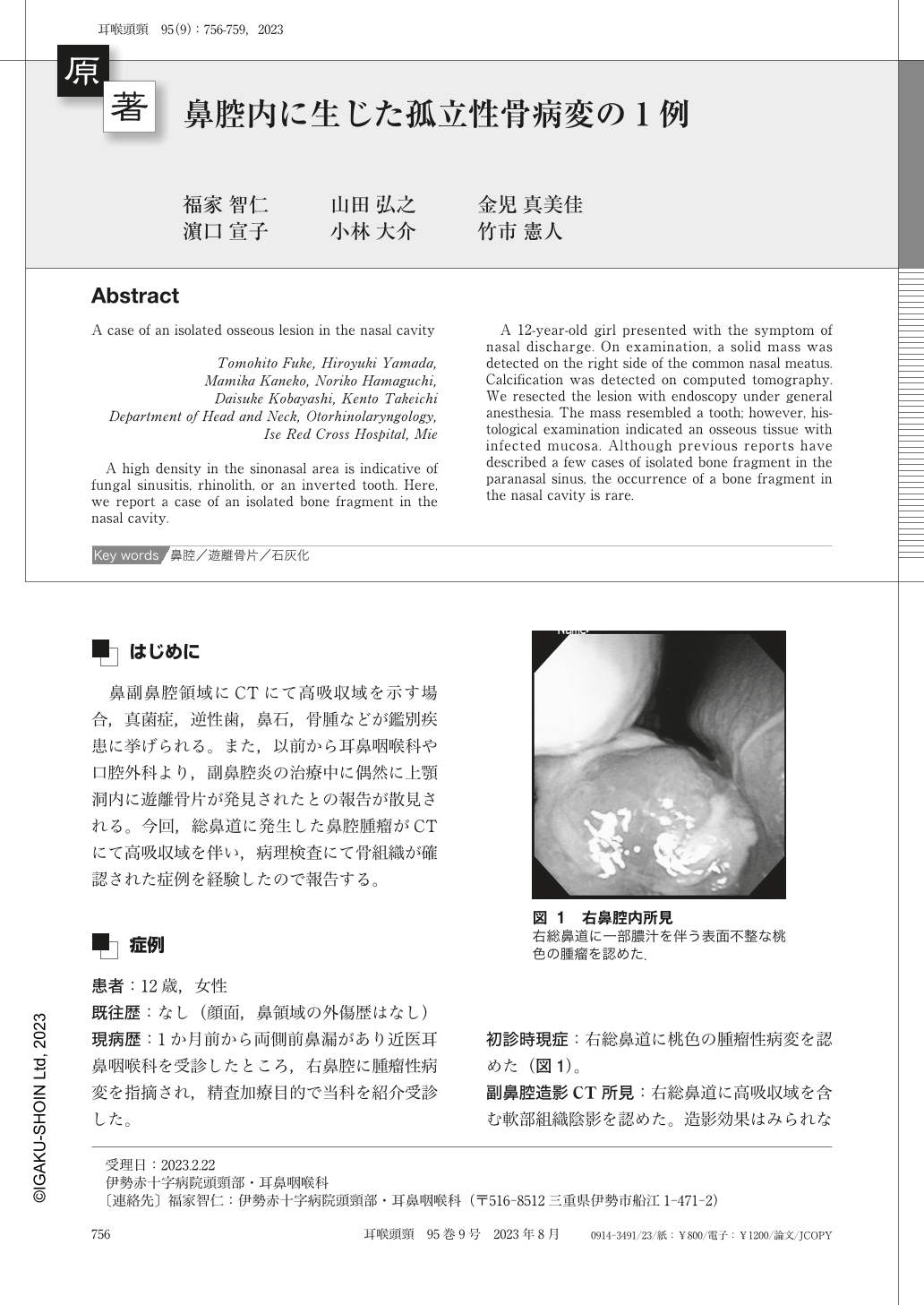
鼻副鼻腔領域にCTにて高吸収域を示す場合,真菌症,逆性歯,鼻石,骨腫などが鑑別疾患に挙げられる。また,以前から耳鼻咽喉科や口腔外科より,副鼻腔炎の治療中に偶然に上顎洞内に遊離骨片が発見されたとの報告が散見される。今回,総鼻道に発生した鼻腔腫瘤がCTにて高吸収域を伴い,病理検査にて骨組織が確認された症例を経験したので報告する。
A high density in the sinonasal area is indicative of fungal sinusitis, rhinolith, or an inverted tooth. Here, we report a case of an isolated bone fragment in the nasal cavity.
A 12-year-old girl presented with the symptom of nasal discharge. On examination, a solid mass was detected on the right side of the common nasal meatus. Calcification was detected on computed tomography. We resected the lesion with endoscopy under general anesthesia. The mass resembled a tooth; however, histological examination indicated an osseous tissue with infected mucosa. Although previous reports have described a few cases of isolated bone fragment in the paranasal sinus, the occurrence of a bone fragment in the nasal cavity is rare.

Copyright © 2023, Igaku-Shoin Ltd. All rights reserved.


