Japanese
English
原著
下垂体腫瘍摘出術後に脳膿瘍を形成した蝶形骨洞篩骨洞囊胞の1例
A Case of Paranasal Cyst with Brain Abscess Presenting Visual Loss in 23 Years after Trans-Sphenoid Surgery
藤岡 正人
1
,
五島 史行
1,2
,
國弘 幸伸
1
,
荒木 康智
1
,
深谷 和正
3
,
小川 郁
1
Masato Fujioka
1
1慶應義塾大学医学部耳鼻咽喉科学教室
2日野市立病院耳鼻咽喉科
3佐野厚生病院耳鼻咽喉科
1Department of Otolaryngology,Keio University School of Medicine
pp.867-870
発行日 2004年11月20日
Published Date 2004/11/20
DOI https://doi.org/10.11477/mf.1411100682
- 有料閲覧
- Abstract 文献概要
- 1ページ目 Look Inside
I.はじめに
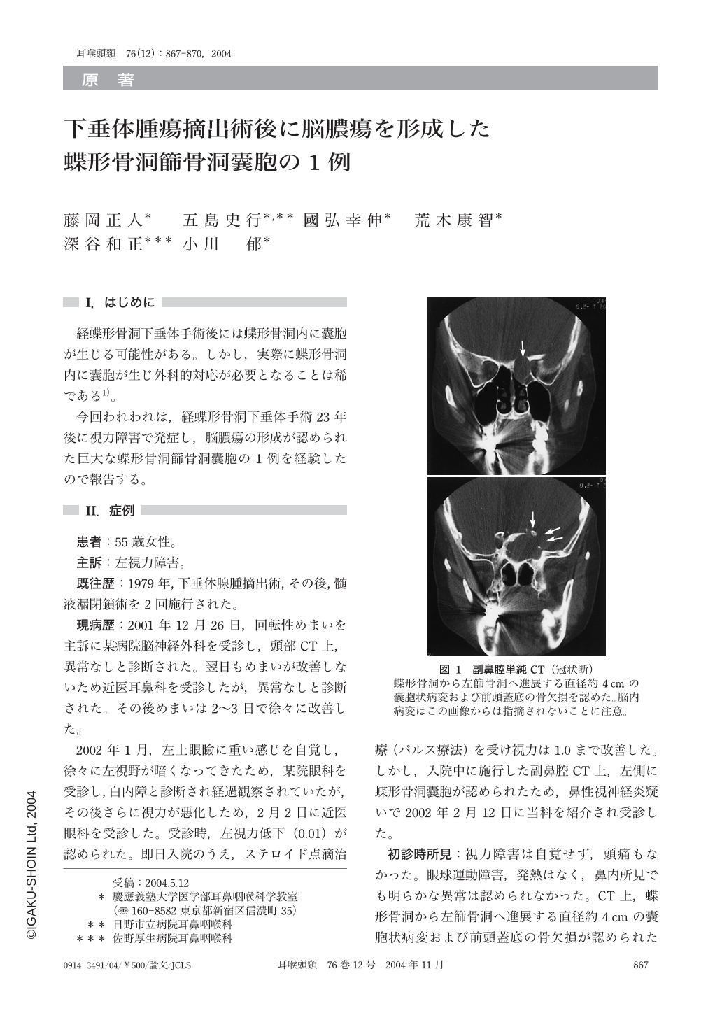
経蝶形骨洞下垂体手術後には蝶形骨洞内に囊胞が生じる可能性がある。しかし,実際に蝶形骨洞内に囊胞が生じ外科的対応が必要となることは稀である1)。
今回われわれは,経蝶形骨洞下垂体手術23年後に視力障害で発症し,脳膿瘍の形成が認められた巨大な蝶形骨洞篩骨洞囊胞の1例を経験したので報告する。
A patient received steroid therapy for visual disturbance,and her visual acuity was recovered.
Then a small defect in the anterior skull base was found on plain brain CT,and a paranasal cyst was found on sinus CT. In addition a brain abscess was revealed by head MRI.
The patient underwent trans-sphenoid surgery for hypophyeal tumor 23 years ago. Optic neuritis caused by rhinogenic origin should be considered at the first visit. We now understand that area of CT scan as well as choice of imaging modality is important for the correct diagnosis.

Copyright © 2004, Igaku-Shoin Ltd. All rights reserved.


