Japanese
English
特集 第20回臨床眼科学会講演集(その3)
篩骨および蝶形骨洞拡大による失明例
A Case of Chronic Retrobulbar Neuritis Secondary to Posterior Accessory Sinusitis
小原 実
1
Minoru Ohara
1
1神戸大学医学部眼科学教室
1Department of Ophthalmology, Kobe University, School of Medicine
pp.523-527
発行日 1967年4月15日
Published Date 1967/4/15
DOI https://doi.org/10.11477/mf.1410203634
- 有料閲覧
PPV購入案内
1,320円 (1,200円+税10%) こちらは、523ページから527ページの文献です。
PPV(ペイ・パー・ビュー)とは、論文(記事)単位で購入・閲覧ができるサービスです。
購入には医書.jp本会員登録が必要です。
会員登録完了後に改めて上記「購入サイトへ」を選択してください。
または「購入サイトへ」選択後に「購入ログイン」、「新規会員登録」の順に進んでください。
会員登録について詳しくはをご確認ください。
購入には医書.jp本会員登録が必要です。
会員登録完了後に改めて上記「購入サイトへ」を選択してください。
または「購入サイトへ」選択後に「購入ログイン」、「新規会員登録」の順に進んでください。
会員登録について詳しくはをご確認ください。
- Abstract 文献概要
- 1ページ目 Look Inside
I.緒言
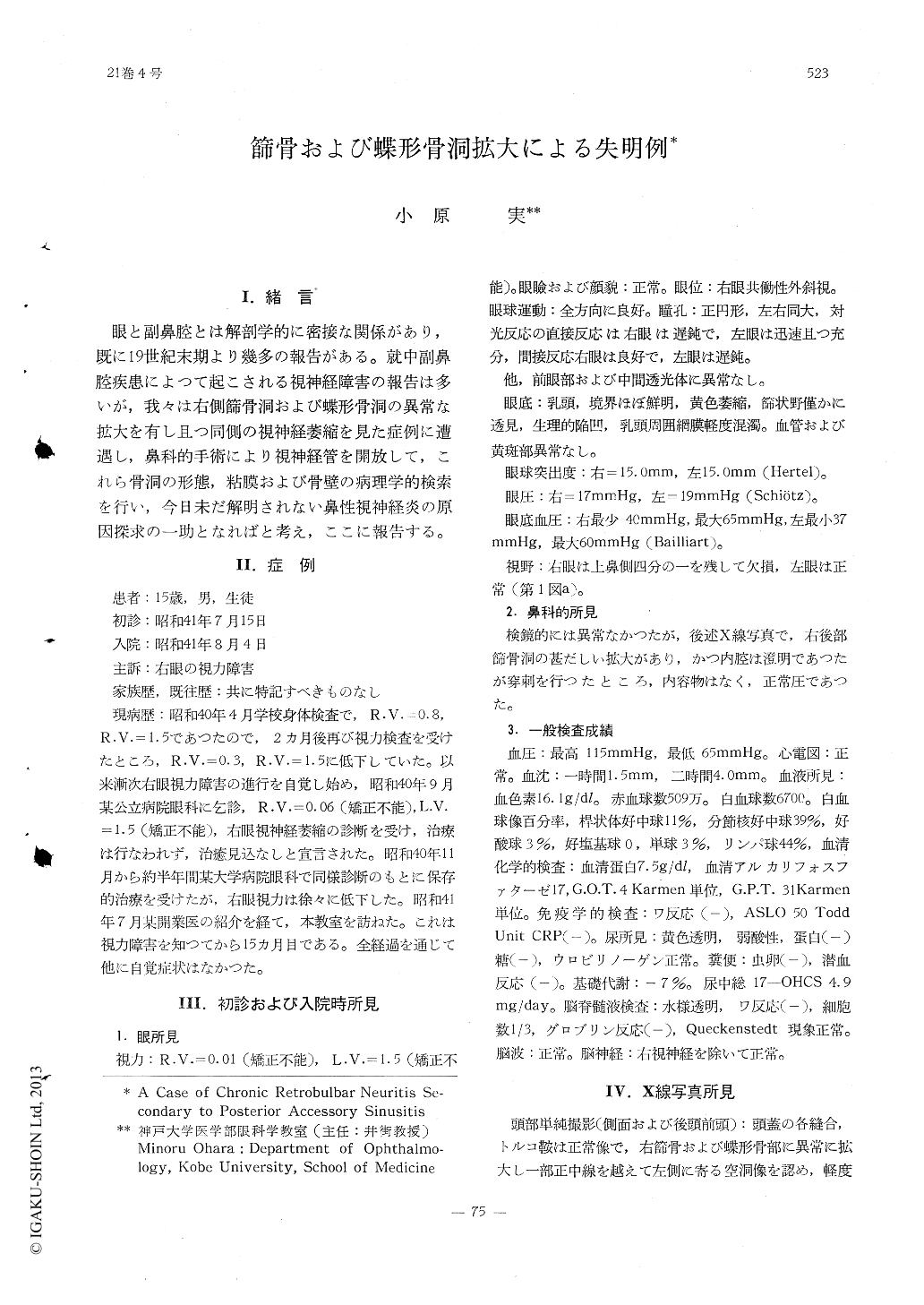
眼と副鼻腔とは解剖学的に密接な関係があり,既に19世紀末期より幾多の報告がある。就中副鼻腔疾患によつて起こされる視神経障害の報告は多いが,我々は右側篩骨洞および蝶形骨洞の異常な拡大を有し且つ同側の視神経萎縮を見た症例に遭遇し,鼻科的手術により視神経管を開放して,これら骨洞の形態,粘膜および骨壁の病理学的検索を行い,今日未だ解明されない鼻性視神経炎の原因探求の一助となればと考え,ここに報告する。
A 15-year-old boy was examined because of decreased visual acuity of right eye to 0.01 (n.c.) since one year. Ophthalmological exami-nation had revealed post-inflammatory optic nerve atrophy and visual field defect of right side. By X-ray examination extraordinary en-largement of right ethmoid and sphenoid sinus was detected. Opening of the optic canal by transnasal approach was performed. Histologi-cal finding of mucous membrane of the sinus showed hypertrophic inflammatior.
Copyright © 1967, Igaku-Shoin Ltd. All rights reserved.