Japanese
English
- 有料閲覧
PPV購入案内
1,320円 (1,200円+税10%) こちらは、543ページから547ページの文献です。購入には医書.jp本会員登録が必要です。
会員登録完了後に改めて上記「購入サイトへ」を選択してください。
または「購入サイトへ」選択後に「購入ログイン」、「新規会員登録」の順に進んでください。
会員登録について詳しくはをご確認ください。
- Abstract 文献概要
- 1ページ目 Look Inside
I.はじめに
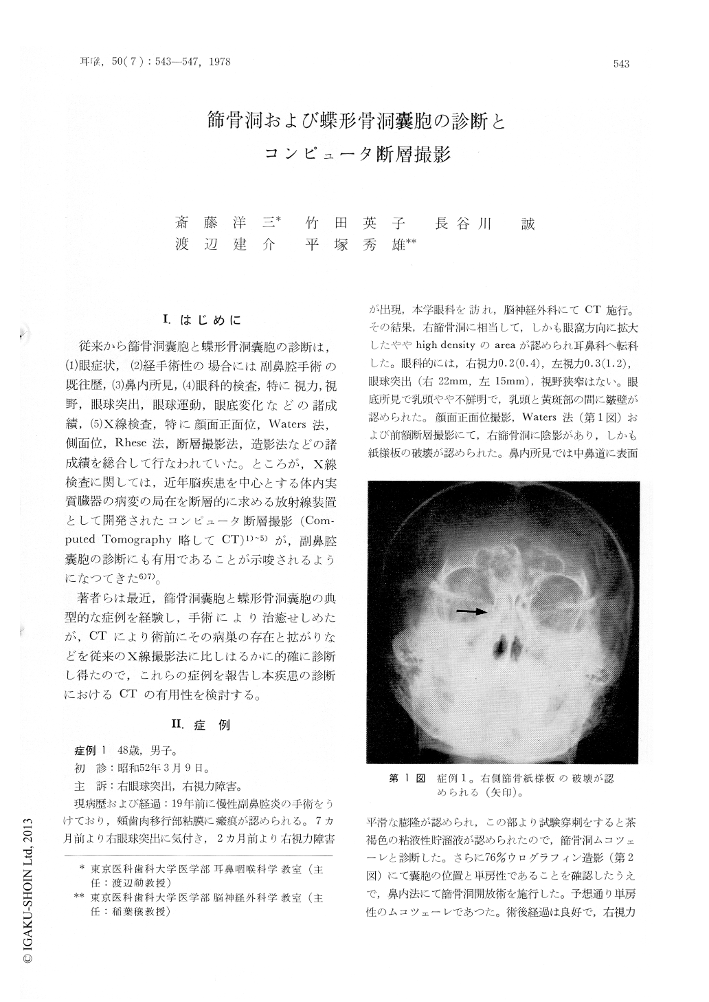
従来から篩骨洞嚢胞と蝶形骨洞嚢胞の診断は,(1)眼症状,②経手術性の場合には副鼻腔手術の既往歴,(3)鼻内所見,(4)眼科的検査,特に視力,視野,眼球突出,眼球運動,眼底変化などの諸成績,(5)X線検査,特に顔面正面位,Waters法,側面位,Rhesc法,断層撮影法,造影法などの諸成績を総合して行なわれていた。ところが,X線検査に関しては,近年脳疾患を中心とする体内実質臓器の病変の局在を断層的に求める放射線装置として開発されたコンピュータ断層撮影(Computed Tomography略してCT)1)〜5)が,副鼻腔嚢胞の診断にも有用であることが示唆されるようになつてきた6)7)。
著者らは最近,篩骨洞嚢胞と蝶形骨洞嚢胞の典型的な症例を経験し,手術により治癒せしめたが,CTにより術前にその病巣の存在と拡がりなどを従来のX線撮影法に比しはるかに的確に診断し得たので,これらの症例を報告し本疾患の診断におけるCTの有用性を検討する。
Computed tomography (CT) was used for the diagnosis of an ethmoidal and a sphenoidal cyst in two cases. These cysts presented slightly high density or isodensity in the preoperative CT scanning, and they protruded into the orbital cavity. In the postoperative CT scanning, the ethmoidal and sphenoidal area in these cases showed low density and the protrusion into the orbital cavity disappeared. CT showed the outline and the content of the cysts clearer than the conventional x-ray examinations.
CT is an useful examination for the diagnosis of the cysts in the ethmoidal and sphenoidal regions.
Copyright © 1978, Igaku-Shoin Ltd. All rights reserved.