Japanese
English
原著
視力障害を伴った巨大な蝶形骨洞囊胞の1例
A Case of Sphenoid Sinus Pyocele with Bilateral Visual Disturbance
竹内 彩子
1
,
武田 靖志
2
,
岡野 光博
2
,
福島 邦博
2
,
小川 晃弘
3
,
菅田 研一
1
,
赤木 成子
1
Ayako Takeuchi
1
1岡山赤十字病院耳鼻咽喉科
2岡山大学大学院医歯学総合研究所 耳鼻咽喉・頭頸部外科学
3姫路聖マリア病院耳鼻咽喉科
1Department of Otorhinolaryngology,Okayama Red Cross Hospiral
pp.60-63
発行日 2005年1月20日
Published Date 2005/1/20
DOI https://doi.org/10.11477/mf.1411100065
- 有料閲覧
- Abstract 文献概要
- 1ページ目 Look Inside
- サイト内被引用 Cited by
I.はじめに
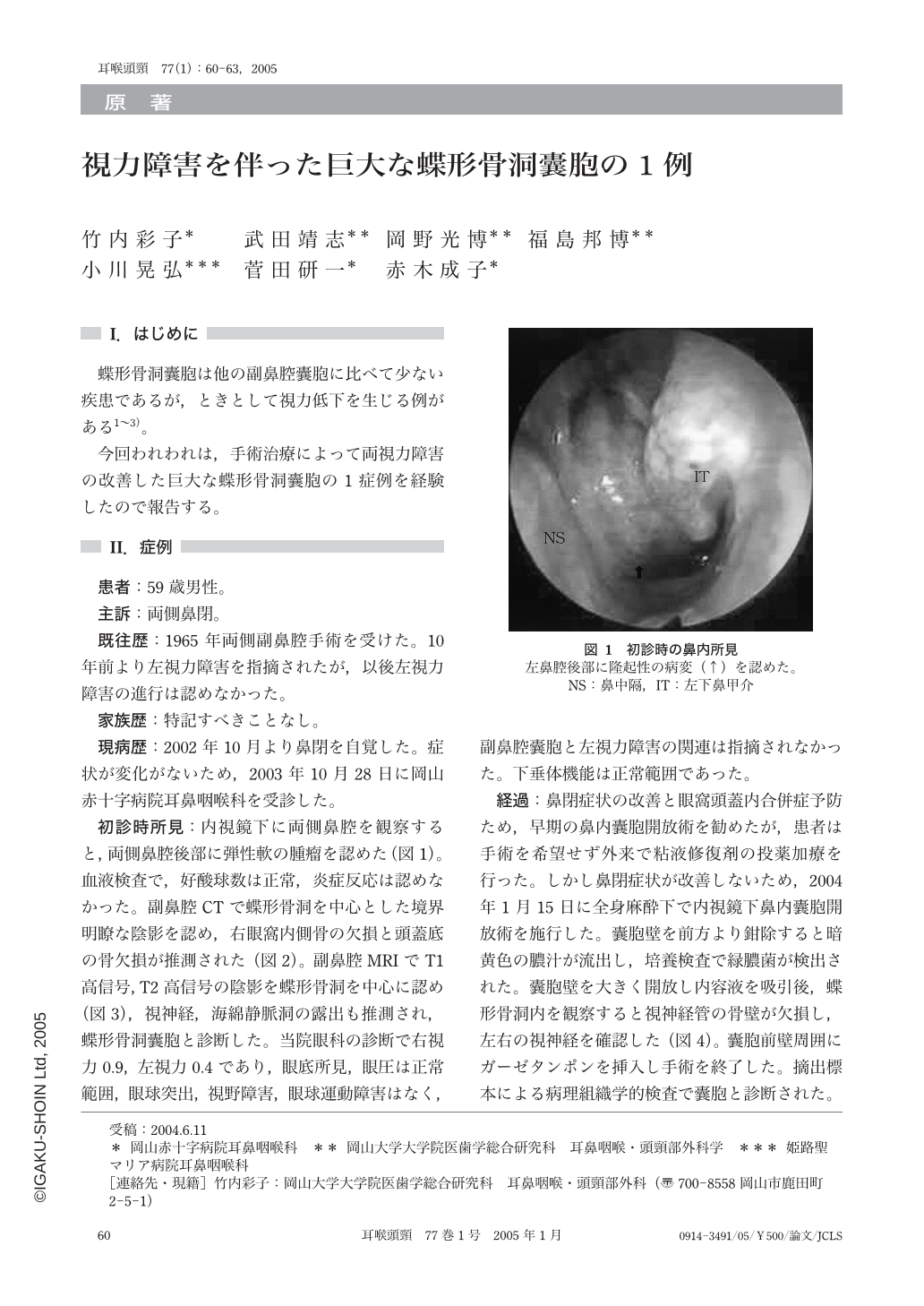
蝶形骨洞囊胞は他の副鼻腔囊胞に比べて少ない疾患であるが,ときとして視力低下を生じる例がある1~3)。
今回われわれは,手術治療によって両視力障害の改善した巨大な蝶形骨洞囊胞の1症例を経験したので報告する。
A 59-year-old man suffered from nasal obstruction in October 2002. He had an operation for bilateral sinectomy 40 years ago. A cystic shadow was recognized in the sphenoidal sinuses by CT scan and MRI. We performed an endonasal endoscopic operation and the wall of the pyocele was removed. His vision recovered one week after the operation.
No bilateral visual disturbance recurred up to the present time.

Copyright © 2005, Igaku-Shoin Ltd. All rights reserved.


