Japanese
English
- 有料閲覧
- Abstract 文献概要
- 1ページ目 Look Inside
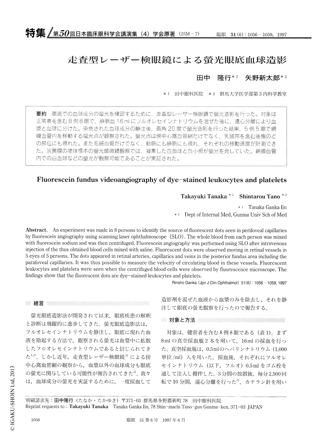
(25M-7) 眼底での血球成分の螢光を確認するために,走査型レーザー検眼鏡で螢光造影を行った。対象は正常者を含む8例8眼で,静脈血16mlにフルオレセインナトリウムを混ぜた後に,遠心分離により血漿と血球に分けた。染色された血球成分の静注後,画角20度で螢光造影を行った結果,5例5眼で網膜血管内を移動ずる螢光点が観察された。螢光点は傍中心窩血管網だけでなく,乳頭部を含む後極のどの部位にも現れた。また毛細血管だけでなく,動脈にも静脈にも現れ,それぞれの移動速度が計測できた。淡黄膜の塗抹標本の螢光顕微鏡観察では,凝集した白血球と血小板が螢光を発していた。網膜血管内での白血球などの螢光が観察可能であることが実証された。
An experiment was made in 8 persons to identify the source of fluorescent dots seen in perifoveal capillaries by fluorescein angiography using scanning laser ophthalmoscope (SLO). The whole blood from each person was mixed with fluorescein sodium and was then centrifuged. Fluorescein angiography was performed using SLO after intravenous injection of the thus obtained blood cells mixed with saline. Fluorescent dots were observed moving in retinal vessels in 5 eyes of 5 persons. The dots appeared in retinal arteries, capillaries and veins in the posterior fundus area including the parafoveal capillaries. It was thus possible to measure the velocity of circulating blood in these vessels. Fluorescent leukocytes and platelets were seen when the centrifuged blood cells were observed by fluorescence microscope. The findings show that the fluorescent dots are dye-stained leukocytes and platelets.

Copyright © 1997, Igaku-Shoin Ltd. All rights reserved.


