Japanese
English
- 有料閲覧
- Abstract 文献概要
- 1ページ目 Look Inside
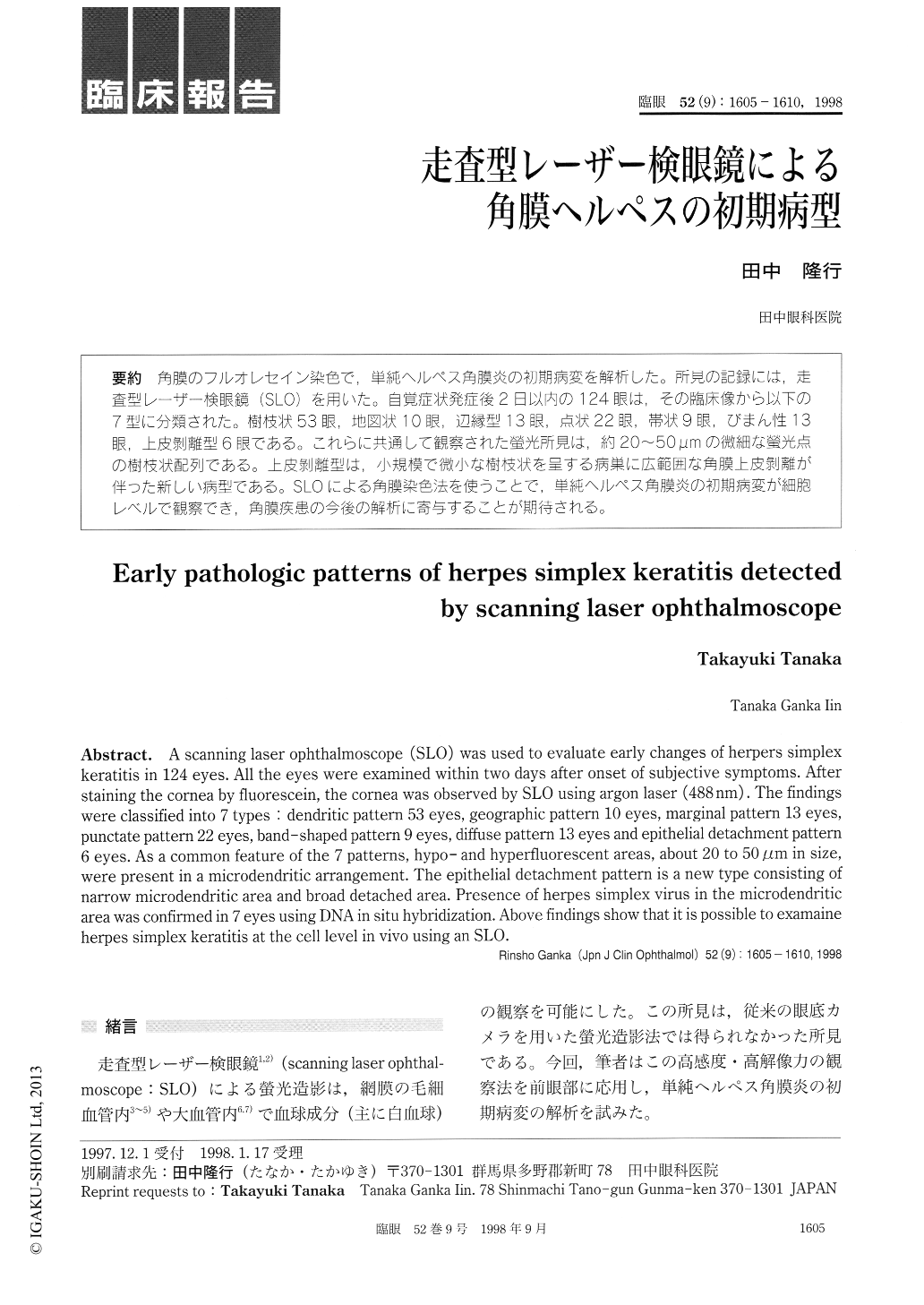
角膜のフルオレセイン染色で,単純ヘルペス角膜炎の初期病変を解析した。所見の記録には,走査型レーザー検眼鏡(SLO)を用いた。自覚症状発症後2日以内の124眼は,その臨床像から以下の7型に分類された。樹枝状53眼,地図状10眼,辺縁型13眼,点状22眼,帯状9眼,びまん性13眼,上皮剥離型6眼である。これらに共通して観察された螢光所見は,約20〜50μmの微細な螢光点の樹枝状配列である。上皮剥離型は,小規模で微小な樹枝状を呈する病巣に広範囲な角膜上皮剥離が伴った新しい病型である。SLOによる角膜染色法を使うことで,単純ヘルペス角膜炎の初期病変が細胞レベルで観察でき,角膜疾患の今後の解析に寄与することが期待される。
A scanning laser ophthalmoscope (SLO) was used to evaluate early changes of herpers simplex keratitis in 124 eyes. All the eyes were examined within two days after onset of subjective symptoms. After staining the cornea by fluorescein, the cornea was observed by SLO using argon laser (488 nm) . The findings were classified into 7 types : dendritic pattern 53 eyes, geographic pattern 10 eyes, marginal pattern 13 eyes, punctate pattern 22 eyes, band-shaped pattern 9 eyes, diffuse pattern 13 eyes and epithelial detachment pattern 6 eyes. As a common feature of the 7 patterns, hypo- and hyperfluorescent areas, about 20 to 50 gm in size, were present in a microdendritic arrangement. The epithelial detachment pattern is a new type consisting of narrow microdendritic area and broad detached area. Presence of herpes simplex virus in the microdendritic area was confirmed in 7 eyes using DNA in situ hybridization. Above findings show that it is possible to examaine herpes simplex keratitis at the cell level in vivo using an SLO.

Copyright © 1998, Igaku-Shoin Ltd. All rights reserved.


