Japanese
English
- 有料閲覧
- Abstract 文献概要
- 1ページ目 Look Inside
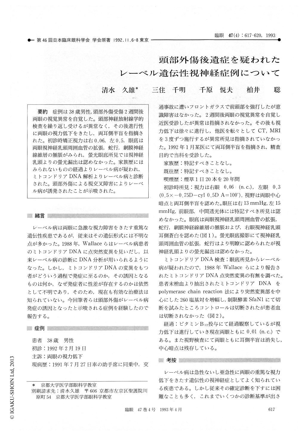
症例は38歳男性,頭部外傷受傷2週間後両眼の視覚異常を自覚した。頭部神経放射線学的検査を繰り返し受けるが異常なく,その後進行性に両眼の視力低下をきたし,両耳側半盲を指摘された。初診時矯正視力は右0.06,左0.5,眼底は両眼視神経乳頭周囲血管の拡張、蛇行,網膜神経線維層の腫脹がみられ,螢光眼底所見では視神経乳頭よりの螢光漏出は認めなかった。家族歴にはみられないものの経過よりレーベル病が疑われ,ミトコンドリアDNA解析よりレーベル病と診断された。頭部外傷による視交叉障害によりレーベル病が誘発されたことが示唆された。
A 38-year-old male presented with bilateral loss of visual acuity 2 weeks after closed head injury. There was no loss of consciousness after the trauma. The corrected visual acuity was 0.06 right and 0.5 left. He showed bilateral central scotoma and bitemporal hemianopia. Funduscopy showed peripapillary microangiopathy and swelling of nerve fiber layer simulating Leber's hereditary optic neuropathy. Fluorescein angiography showed normal findings. The family history was negative. Mitochondrial DNA studies showed 11778 muta-tion. Bitemporal hemianopia disappeared 6 months later leaving dense bilateral central scotoma. The final visual acuity was 0.01 in each eye. The clini-cal course suggested that chiasmal dysfunctions secondary to head injury triggered manifestation of Leber's hereditary optic neuropathy.

Copyright © 1993, Igaku-Shoin Ltd. All rights reserved.


