Japanese
English
- 有料閲覧
- Abstract 文献概要
- 1ページ目 Look Inside
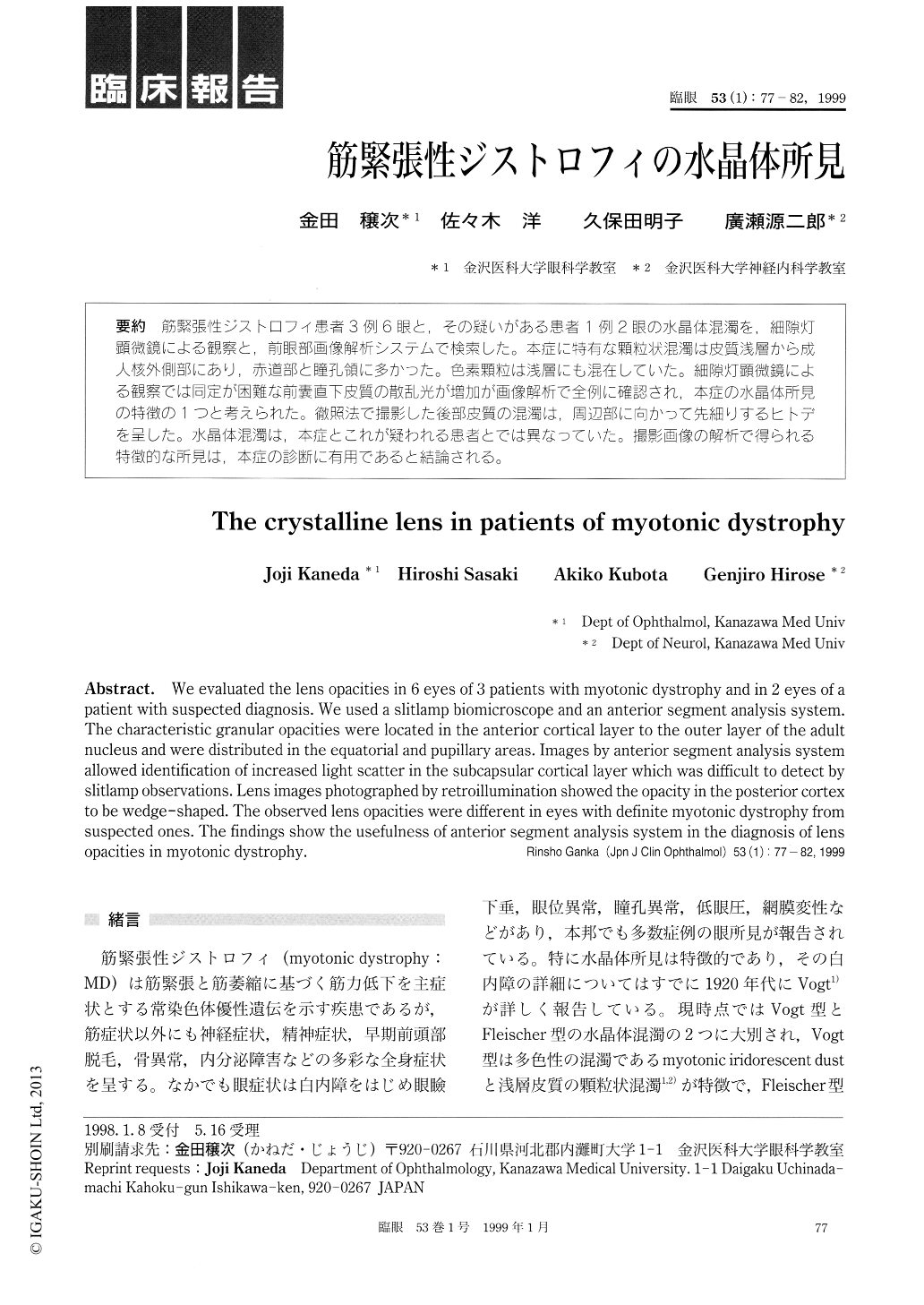
筋緊張性ジストロフィ患者3例6眼と、その疑いがある患者1例2眼の水晶体混濁を,細隙灯顕微鏡による観察と,前眼部画像解析システムで検索した。本症に特有な顆粒状混濁は皮質浅層から成人核外側部にあり,赤道部と瞳孔領に多かった。色素顆粒は浅層にも混在していた。細隙灯顕微鏡による観察では同定が困難な前嚢直下皮質の散乱光が増加が画像解析で全例に確認され,本症の水晶体所見の特徴の1つと考えられた。徹照法で撮影した後部皮質の混濁は,周辺部に向かって先細りするヒトデを呈した。水晶体混濁は,本症とこれが疑われる患者とでは異なっていた。撮影画像の解析で得られる特徴的な所見は,本症の診断に有用であると結論される。
We evaluated the lens opacities in 6 eyes of 3 patients with myotonic dystrophy and in 2 eyes of a patient with suspected diagnosis. We used a slitlamp biomicroscope and an anterior segment analysis system. The characteristic granular opacities were located in the anterior cortical layer to the outer layer of the adult nucleus and were distributed in the equatorial and pupillary areas. Images by anterior segment analysis system allowed identification of increased light scatter in the subcapsular cortical layer which was difficult to detect by slitlamp observations. Lens images photographed by retroillumination showed the opacity in the posterior cortex to be wedge-shaped. The observed lens opacities were different in eyes with definite myotonic dystrophy from suspected ones. The findings show the usefulness of anterior segment analysis system in the diagnosis of lens opacities in myotonic dystrophy.

Copyright © 1999, Igaku-Shoin Ltd. All rights reserved.


