Japanese
English
- 有料閲覧
- Abstract 文献概要
- 1ページ目 Look Inside
- サイト内被引用 Cited by
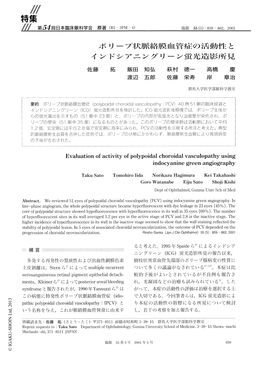
ポリープ状脈絡膜血管症(polypoidaL choroidal vasculopathy:PCV)40例51眼の臨床経過とインドシアニングリーン(ICG)蛍光造影所見を検討した。ICG蛍光造影後期像では,ポリープ全体からの蛍光漏出を示すもの(51眼中23眼)と,ポリープの内部が低蛍光となり血管壁が染色され,ポリープの壁染(51眼中35眼)になるものとがあった。このポリープの壁染数は活動期において平均1.2個,安定期には平均2.8個で安定期に高率にみられ,PCVの活動性を示唆ずる所見と考えた。典型的脈絡膜新生血管を合併した症例では,ポリープの状態にかかわらず,脈絡膜新生血管により黄斑病変の予後が左右された。
We reviewed 51 eyes of polypoidal choroidal vasculopathy (PCV) using indocyanine green angiography. Inlate-phase angiogram, the whole polypoidal structure became hyperfluorescent with dye leakage in 23 eyes (45%). Thecore of polypoidal structure showed hypofluorescence with hyperfluorescence in its wall in 35 eyes (69%). The numberof hyperfluorescent sites in its wall averaged 1.2 per eye in the active stage of PCV and 2.8 in the inactive stage. Thehigher incidence of hyperfluorescence in its wall in the inactive stage seemed to show that the wall staining reflected the stability of polypoidal lesion. In 5 eyes of associated choroidal neovascularization, the outcome of PCV depended on theprogression of choroidal neovascularization.

Copyright © 2001, Igaku-Shoin Ltd. All rights reserved.


