Japanese
English
- 有料閲覧
- Abstract 文献概要
- 1ページ目 Look Inside
- 参考文献 Reference
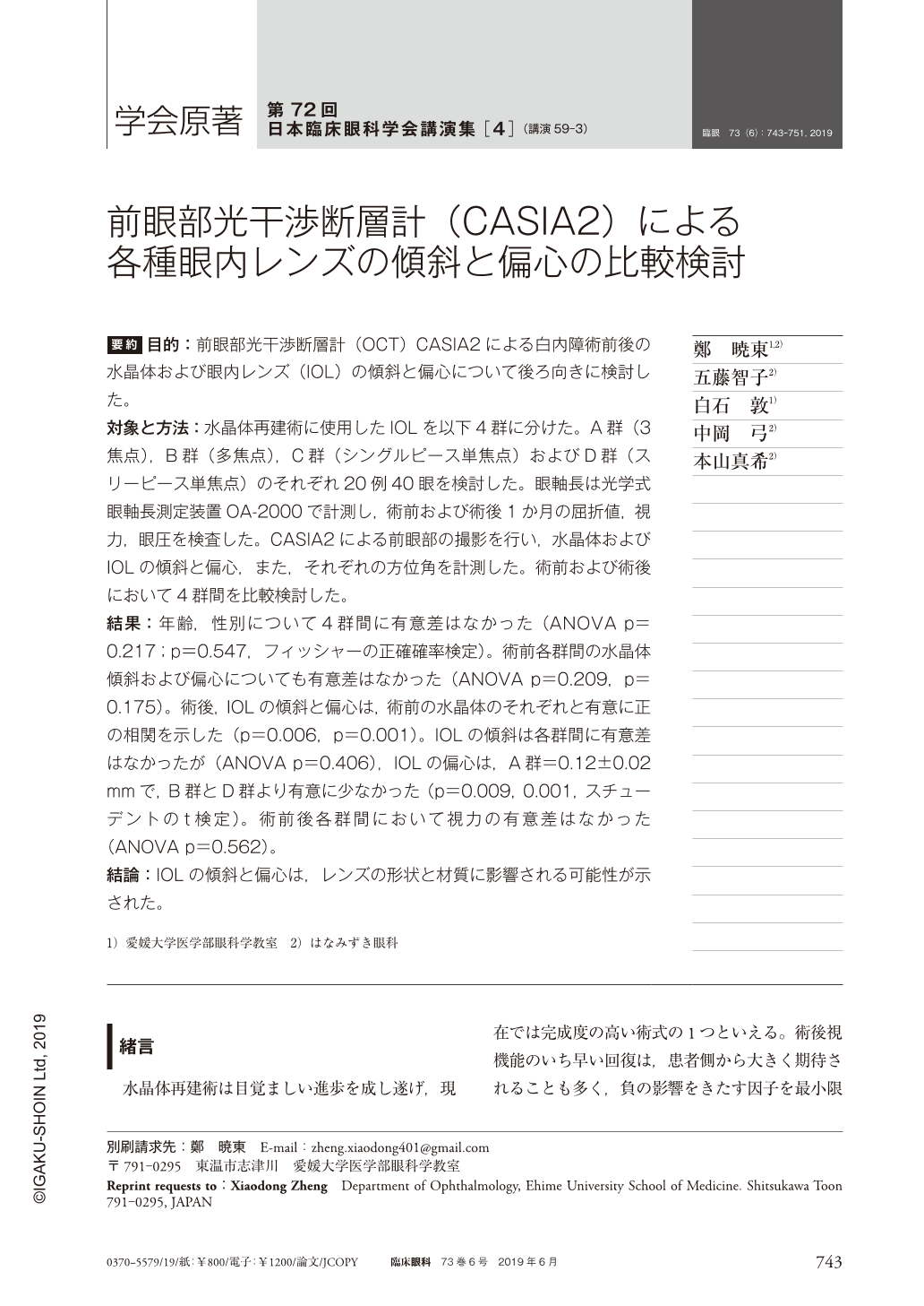
要約 目的:前眼部光干渉断層計(OCT)CASIA2による白内障術前後の水晶体および眼内レンズ(IOL)の傾斜と偏心について後ろ向きに検討した。
対象と方法:水晶体再建術に使用したIOLを以下4群に分けた。A群(3焦点),B群(多焦点),C群(シングルピース単焦点)およびD群(スリーピース単焦点)のそれぞれ20例40眼を検討した。眼軸長は光学式眼軸長測定装置OA-2000で計測し,術前および術後1か月の屈折値,視力,眼圧を検査した。CASIA2による前眼部の撮影を行い,水晶体およびIOLの傾斜と偏心,また,それぞれの方位角を計測した。術前および術後において4群間を比較検討した。
結果:年齢,性別について4群間に有意差はなかった(ANOVA p=0.217;p=0.547,フィッシャーの正確確率検定)。術前各群間の水晶体傾斜および偏心についても有意差はなかった(ANOVA p=0.209,p=0.175)。術後,IOLの傾斜と偏心は,術前の水晶体のそれぞれと有意に正の相関を示した(p=0.006,p=0.001)。IOLの傾斜は各群間に有意差はなかったが(ANOVA p=0.406),IOLの偏心は,A群=0.12±0.02mmで,B群とD群より有意に少なかった(p=0.009,0.001,スチューデントのt検定)。術前後各群間において視力の有意差はなかった(ANOVA p=0.562)。
結論:IOLの傾斜と偏心は,レンズの形状と材質に影響される可能性が示された。
Abstract Purpose:To retrospectively investigate the tilt and decentration of crystalline lens and intraocular lens(IOL)using a second-generation anterior segment optical coherence tomography, CASIA2.
Methods:Patients underwent cataract surgery in Hanamizuki Eye Clinic were studied. Forty eyes of 20 patients each in the following 4 different types of IOL groups were investigated. Group A, trifocal, Finevision(PhysIOL), Group B, multifocal, ZMB(AMO), Group C, single-piece, monofocal, ZCB00V(AMO)and, Group D, three-piece, monofocal, AR40e(AMO). The axial length was measured by OA-2000(Tomey). Refractive power and visual acuity and IOP were measured before and 1 month after surgery. CASIA2(Tomey)was used to examine all patients before and 1 month after surgery. A built-in soft was used to study the tilt and decentration of the crystalline lens and IOL among groups.
Results:There was no significant difference in age and gender among groups(ANOVA p=0.217;p=0.547, Fisher's exact test). There were no significant differences in the tilt and decentration of crystalline lens among groups before surgery(ANOVA p=0.209, p=0.175). The tilt and decentration of IOL were significantly correlated with crystalline lens(p=0.006;p=0.001;Spearman coefficient). IOL had similar amount of tilt among groups. IOL decentration of Group A was 0.12±0.02 mm, significantly smaller than that of Groups B or D(p=0.009, p=0.001, Student t-test). No significant difference was noted in the visual acuity among groups before and after surgery.
Conclusions:The structure and material of IOL may affect its position after surgery.

Copyright © 2019, Igaku-Shoin Ltd. All rights reserved.


