Japanese
English
- 有料閲覧
- Abstract 文献概要
- 1ページ目 Look Inside
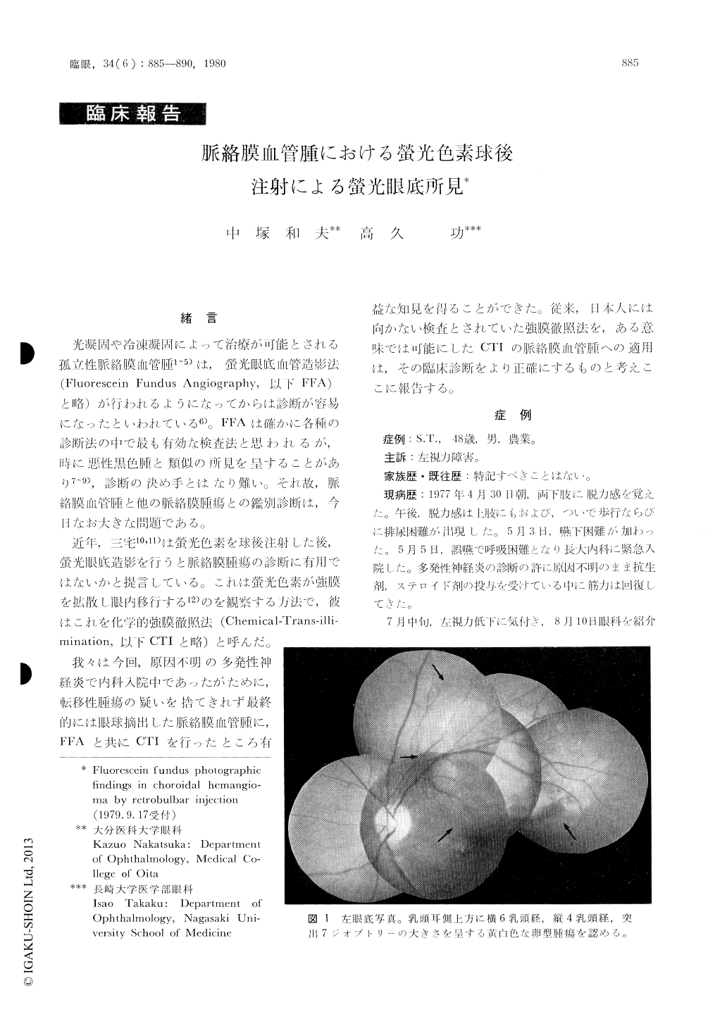
48歳の男子に認めた孤立性脈絡膜血管腫に螢光色素球後注射による螢光眼底造影法(CTI)を行い通常の螢光眼底血管造影法(FFA)と比較検討した。
1) CTIの初期像は,び漫性雲状の螢光を呈すがこれは螢光色素の血管流入によるFFAの早期像と異なり,腫瘍本体の染色によると推定した。
2) CTIの中期から後期にかけては顆粒状螢光が増加するとともに斑状螢光が出現する。これは色素上皮のwindow defectに加え螢光色素が網膜神経上皮下ならびに神経上皮層の類嚢胞状浮腫に貯留するためと考えられる。
3) CTIの螢光は長時間貯留し,時間の経過につれて不鮮明となる。
CTIの中期以降の螢光像は,FFAの螢光像に近似してくる。このことは両者の過螢光発生機序が途中から同一であることを示唆する。
孤立性脈絡膜血管腫の診断は,今日でも困難な問題である。CTIの実施,とりわけFFAとの併用は診断に寄与するところが大きい検査法と判定した。
A 48-year-old male with choroidal angioma was examined by means of fluorescein angiography after retrobulbar injection of the dye (chemical transil-lumination). The angioma was detected during his admission because of polyneuritis of unidentified etiology.
In the early phase of chemical transillumination, there was diffuse cloudy hyperfluorescence of the tumor area. The hyperfluorescence was due to dye staining of the tumor mass. This feature was dif-ferent from the early-phase fluorescein angiogram through paravenous route, in which the tumor demonstrated vascular fluorescence.

Copyright © 1980, Igaku-Shoin Ltd. All rights reserved.


