Japanese
English
- 有料閲覧
- Abstract 文献概要
- 1ページ目 Look Inside
Ⅰ.緒言
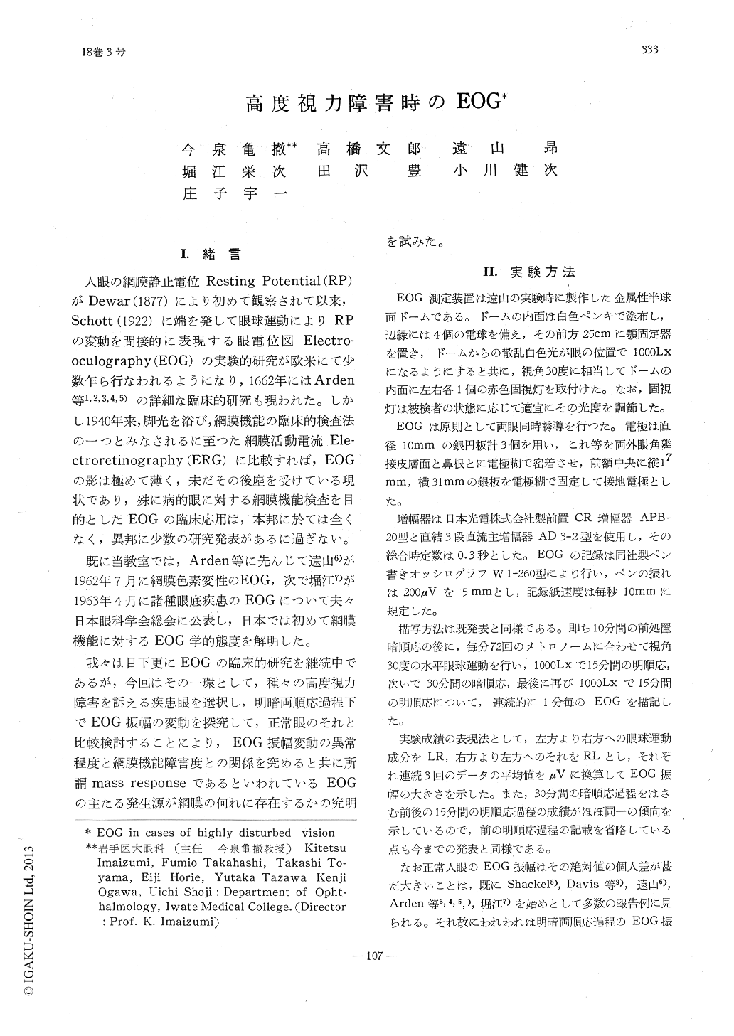
人眼の網膜静止電位Resting Potential (RP)がDewar (1877)により初めて観察されて以来,Schott (1922)に端を発して眼球運動によりRPの変動を間接的に表現する眼電位図Electro—oculography (EOG)の実験的研究が欧米にて少数乍ら行なわれるようになり,1662年にはArden等1,2,3,4,5)の詳細な臨床的研究も現われた。しかし1940年来,脚光を浴び,網膜機能の臨床的検査法の一つとみなされるに至つた網膜活動電流Ele—ctroretinography (ERG)に比較すれば,EOGの影は極めて薄く,未だその後塵を受けている現状であり,殊に病的眼に対する網膜機能検査を目的としたEOGの臨床応用は,本邦に於ては全くなく,異邦に少数の研究発表があるに過ぎない。
既に当教室では,Arden等に先んじて遠山6)が1962年7月に網膜色素変性のEOG,次で堀江7)が1963年4月に諸種眼底疾患のEOGについて夫々日本眼科学会総会に公表し,日本では初めて網膜機能に対するEOG学的態度を解明した。
The electro-oculograms (EOG) in various ocular diseases accompanied by serious visual disturbances were compared with those of normal eyes. The amplitudes of EOGs were determined by recording the changes in pote-ntials which occurred when the eyes were rotated 30 degrees.
The results obtained were as follows:1) The EOG-time curves in normal eyes during dark and light adaptation consist of two portions of dark and light process.
The first concave in the EOG-time curve showed a minimum value (dark trough) at 11 min. in dark adaptaiton and the convex part for light adaptation at 1000Lx. had a steep peak at an average of 7min.

Copyright © 1964, Igaku-Shoin Ltd. All rights reserved.


