Japanese
English
- 有料閲覧
- Abstract 文献概要
- 1ページ目 Look Inside
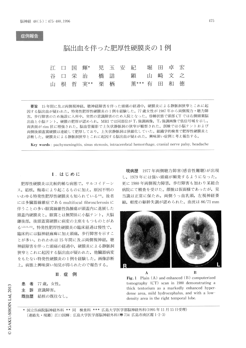
15年間に及ぶ両側視神経,聴神経障害を伴った頭痛の経過中,硬膜炎による静脈洞狭窄とこれに起因する脳出血が疑われた,特発性肥厚性硬膜炎の1例を経験した。77歳女性が1987年から両側視力・聴力障害,歩行障害のため施設に入所中,突然の意識障害のため入院となった。昏睡状態で頭部CTでは右側頭葉脳出血と小脳テント,硬膜の肥厚が認められ,MRIでは同部位がT1強調画像,T2強調画像で低信号域を示し,両表面がrim状に増強された。脳血管撮影で上矢状静脈洞の狭窄が観察された。剖検では小脳テントおよび両側後頭蓋窩硬膜は連続して肥厚しており,上矢状静脈洞は狭細化していた。組織学的検査で肥厚性硬膜炎と診断した。硬膜炎による静脈洞狭窄とこれに起因する脳出血が疑われた,興味深い症例と考え報告する。
A case of cranial hypertrophic pachymeningitis of unknown etiology in a patient with 15-year history of headaches, cranial nerve palsies, and gait distur-bance is reported. A 77-year-old woman was brought to our institute in a coma. CT revealed intracerebral hemorrhage in the right temporal lobe and thickening of the falx and tentorium. Fifteen years previously the patient had undergone CT scanning because of headaches, cranial nerve palsies, and progressive gait disturbance and a thickened tentorium, mild hydrocephalus and edematous change in the right temporal lobe had been reported.

Copyright © 1996, Igaku-Shoin Ltd. All rights reserved.


