Japanese
English
- 有料閲覧
- Abstract 文献概要
- 1ページ目 Look Inside
I.はじめに
硬膜動静脈奇形(以下dAVM)は全頭蓋内AVMの10—15%を占めるが8,11,20),横・S状静脈洞部や海綿静脈洞部に多く,前頭蓋窩dAVMは非常に稀であるとされていた8).しかし近年,報告例が散見され1-3,8-12,16-21),以前よりその頻度は増してきている.
一方,モヤモヤ血管はウイリス動脈輪閉塞症をはじめ,脳動脈硬化症,脳腫瘍など種々の疾患においてもみられることが知られている.中大脳動脈閉塞症においてもモヤモヤ血管を伴う症例が報告されている4-7,13-15)が,これはモヤモヤ病とは本質的には異なった疾患であると考えられている.
The authors report a case of dural arteriovenous mal-formation (dAVM) in the anterior fossa that was associated with an occlusion of the unilateral middle cerebral artery with Moyamoya phenomenon.
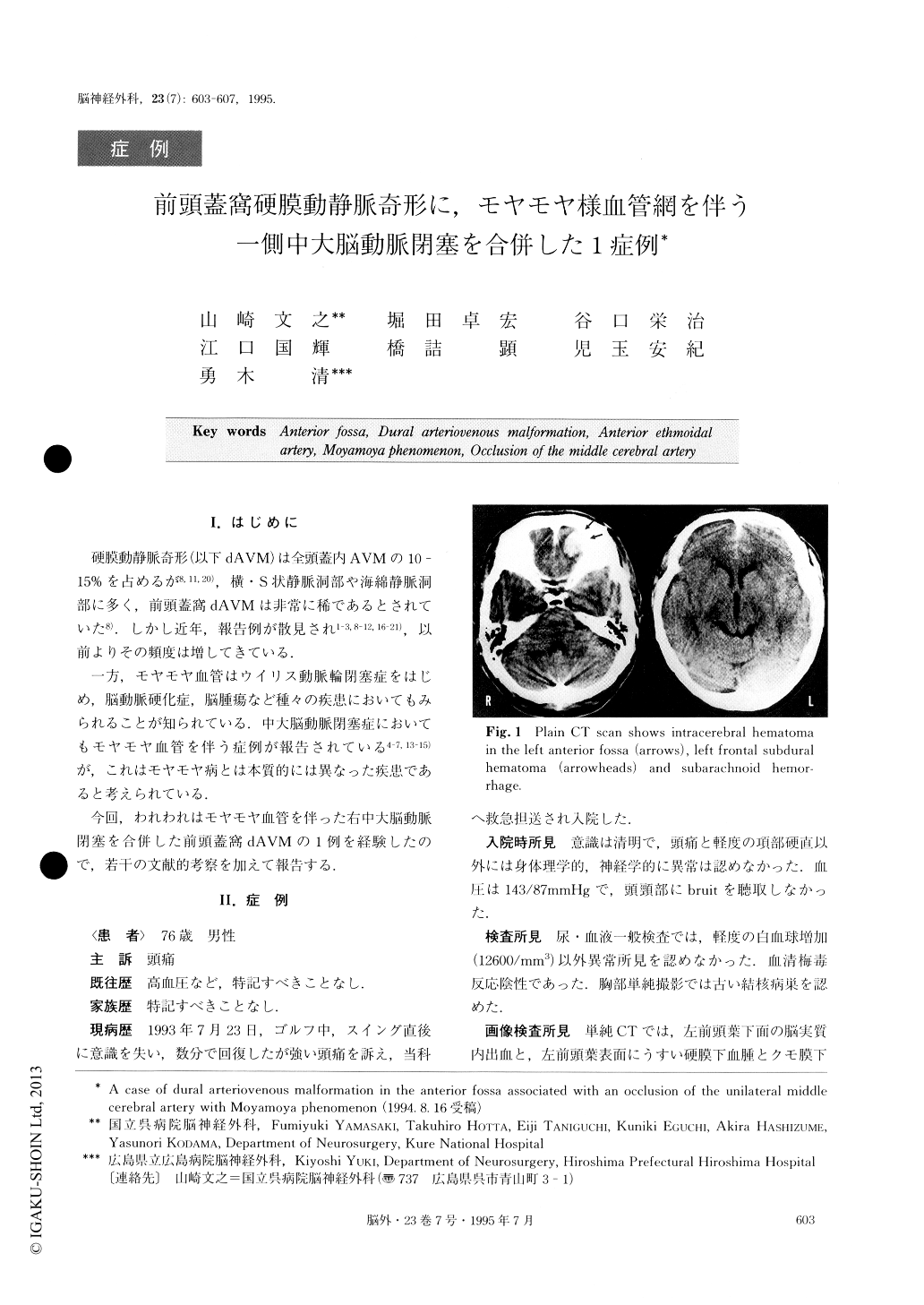
The patient was a 79-year-olcl man referred to our department with an episode of consciousness disturb-ance and headache. Computed tomography scan showed intracerebral hematoma in the left frontal lobe, combined with left frontal subdural hematoma and sub-arachnoid hemorrhage. Angiography revealed dAVM in the left anterior fossa, fed by the bilateral ethmoidal and the bilateral middle meningeal arteries, and drain-ing into the superior and inferior sinuses through the pial veins with vascular sacs. While angiography re-vealed an occlusion of the right middle cerebral artery (M1 portion) good collaterals via leptomeningeal anas-tomosis had developed. T1-weighted magnetic reso-nance (MR) images showed an intracerebral hematoma as a low intensity area, and flow-void areas were in-dicative of vascular sacs. Time-of-flight MR angiogra-phy also clearly showed dAVM in the left anterior fossa.
Co-existence of an occlusion of an intracranial major artery and dAVM in this location has not been re-ported previously and it was speculated that this asso-ciation had occurred by chance.

Copyright © 1995, Igaku-Shoin Ltd. All rights reserved.


