Japanese
English
- 有料閲覧
- Abstract 文献概要
- 1ページ目 Look Inside
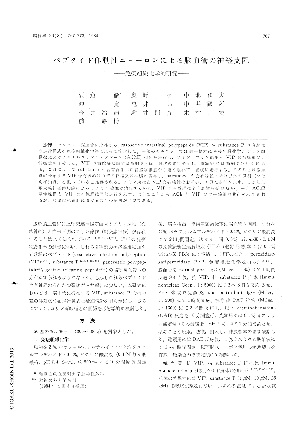
抄録 モルモット脳血管に分布するvasoactive intestinal polypeptide (VIP)やsubstance P含有線維の走行様式を免疫組織化学法によって検討した。一部のモルモットでは同一標本に免疫組織化学とアミン組織螢光又はアセチルコリンエステレース(AChE)染色を施行し,アミン,コリン線維とVIP含有線維の走行様式を比較した。VIP含有線維は血管壁筋細胞と同じ輪状の走行を示し,電顕的には筋細胞の近くに終る。これに反してsubstance P含有線維は血管壁筋細胞から遠く離れて,網状に走行する。このことは脳血管に分布するVIP含有線維は血管の収縮又は拡張に関与し,substance P含有線維はそれ以外の役割(たとえば知覚)を担っていると推察される。アミン線維とVIP含有線維はお互いよく似た走行を示す。しかし上頸交感神経節切除によってアミン線維は消失するのに,VIP含有線維は全く影響を受けない。一方AChE陽性線維とVIP含有線維は同じ走行を示す。以上のことからAChとVIPの同一線維内共存が示唆されるが,なお起始細胞における共存の証明が必要である。
By an immunohistochemical technique, vasoactive polypeptide (VIP)-and substance P-containing nerve fibers are observed in the cerebral bloodvessels. VIP-containing nerve fibers distribute in a spiral pattern, similar to the muscle cell distri-bution pattern. Under an electron microscopic observation, VIP-immunoreactive terminals lie just near to a muscle cell in the inner layer of the adventitia. In contrast, substance P-containing nerve fibers show a meshwork pattern in the outer layer of the adventitia. In combination of acetyl-cholinesterase (AChE) staining and VIP immu-nohistochemistry, AChE-positive and VIP-immu-noreactive nerve fibers reveal almost same dis-tribution in the same specimen. The present data suggest that VIP-containing nerve fibers may play a role of smooth muscle control of the blood vessels, while substance P-containing nerve fibers may not take part in muscle control.

Copyright © 1984, Igaku-Shoin Ltd. All rights reserved.


