Japanese
English
- 有料閲覧
- Abstract 文献概要
- 1ページ目 Look Inside
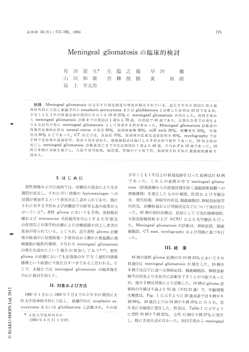
抄録 Meningeal gliomatosisは近年その発生頻度の増加が報告されている。最近2年半の期間に阪大脳神経外科に入院し組織学的にanaplastic astrocytomaまたはglioblastomaと診断した症例は43例であるが,少なくとも1年の経過追跡の期間にそのうち10例23%にmeningeal gliomatosisが発生した。初回手術からmeningeal gliomatosis診断までの期間は1週から78週,中間値で45週であり,長期生存者での発生よりも比較的早期にmeningeal gliomatosisとして再発する例が多かった。Meningeal gliomatosis診断後の特徴的な神経症状はmental statusの異常80%,脳神経麻痺80%,stiff neck 80%,痙攣発作50%,呼吸障害80%などであった。CT所見では,水頭症70%,脳室壁の広範な造影陽性を60%,myelographyでは2例で造影剤の通過障害,陰影欠損を認めた。髄液細胞診は施行した9例全例で陽性であった。10例全例が死亡しmeningeal gliomatosis診断後死亡までの生存期間は1週より46週,平均わずか18週であった。10例中6例に剖検を施行し,大脳半球穹隆部,脳底部,脊髄のクモ膜下腔,脳室壁それぞれに腫瘍細胞播種を認めた。
Ten (23%) patients out of 43 with malignant glioma developed meningeal gliomatosis during the follow up period of at least one year. The duration between the first surgery and diagnosis of meningeal gliomatcsis ranged from one to 78 weeks (median 45 weeks). In younger age group less than 20 years old, 5 (56%) cut of 9 patients had meningeal gliomatosis, and cn the ccntrary the incidence was lower in older age group above 20 years old (5 of 34, 15%). Seven (22%) out of 32 male and 3 (27%) out of 11 female patients developed meningeal gliomatosis. The primary tumor location were frontal lobe in 4 cases (including one bifron-tal tumor), temporal in 2, parieto-occipital in 1, thalamus in 1, midbrain in 1, and cerebellar hemi-sphere in 1, respectively. Histologically, 7 tumors were anaplastic astrocytoma, and 3 were glioblas-toma. The characteristic neurological findings observed during the course of meningeal glioma-tosis were abnormal mental status (80%), cranial nerve palsies (50%), paraplegia (60%), stiff neck (80%), seizure (50%), and respiratory disturbance(80%), CSF cytology was positive in all 9 patients tested. CT scan demonstrated hydrocephalus (70 %), and diffuse contrast enhancement of ventri-cular wall (60%) and basal cistern (10%). In 2 cases, block and irregular filling defect were seen by myelography. Six patients were treated by ir-radiation to the whole brain and/or spine, and 5, by intrathecal chemotherapy with methotrexate, cy-tosine arabinoside and bleomycin. However, all patients died of the tumor one to 46 weeks (me-dian 18 weeks) after the diagnosis of meningeal gliomatosis. Six cases were autopsied, and gross and/or microscopical tumor cell dissemination were seen in the subarachnoid space of the cerebral convexity (83%), ventricular wall (100%), basal cistern (100%), and spinal subarachnoid space (100 %), respectively. Thus, the incidence of meningeal gliomatosis is higher than previously reported, and might be one of the principal causes of the early recurrence and death in patients with malig-nant glioma.

Copyright © 1984, Igaku-Shoin Ltd. All rights reserved.


