Japanese
English
- 有料閲覧
- Abstract 文献概要
- 1ページ目 Look Inside
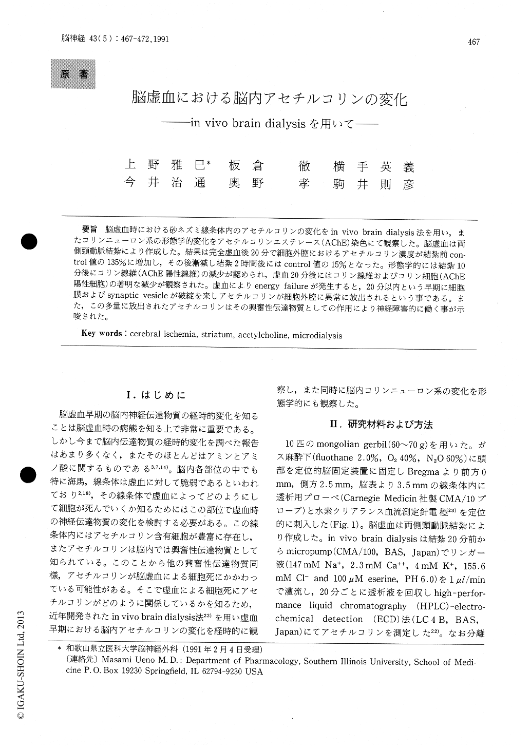
脳虚血時における砂ネズミ線条体内のアセチルコリンの変化をin vivo brain dialysis法を用い,またコリンニューロン系の形態学的変化をアセチルコリンエステレース(AChE)染色にて観察した。脳虚血は両側頸動脈結紮により作成した。結果は完全虚血後20分で細胞外腔におけるアセチルコリン濃度が結紮前con—trol値の135%に増加し,その後漸減し結紮2時間後にはcontrol値の15%となった。形態学的には結紮10分後にコリン線維(AChE陽性線維)の減少が認められ,虚血20分後にはコリン線維およびコリン細胞(AChE陽性細胞)の著明な減少が観察された。虚血によりenergy failureが発生すると,20分以内という早期に細胞膜およびsynaptic vesicleが破綻を来しアセチルコリンが細胞外腔に異常に放出されるという事である。また,この多量に放出されたアセチルコリンはその興奮性伝達物質としての作用により神経障害的に働く事が示唆された。
Neurons in the hippocumpus and striatum are vulnerable to the ischemic insult. In the present study we focused on the vulnerability of acetyl-choline (ACh)-containing neurons in the striatum. To determine the change of ACh content in the striatum, we employed a simple sensive brain mi- crodialysis method with high performance liquid chromatographyelectrochemical detection. Mot.- phological change of cholinergic neurons was stud-ied by ACh-esterase staining during ischemic insult. After bilateral carotid artery occlusion, local cere-bral flow decreased less than 5ml/min/100g in the striatum. Ach content in dialysate for 20min afterthe occlusion was elevated to 135% of the control. ACh content decreased to 15% of the control 2hs after the ischemia. Density of Cholinergic fibers and cell bodies was reduced 10 min after the ischemia and further decreased 20min after the occlusion. Our study suggests that the trasient elevation of ACh content was probably due to diffusion of AChfrom destructed cholinergic terminals after the is-chemic insult. The increase of ACh immediately after arterial occlusion may affect severity of is-chemic damage in the brain by its excitatory effect.

Copyright © 1991, Igaku-Shoin Ltd. All rights reserved.


