Japanese
English
- 有料閲覧
- Abstract 文献概要
- 1ページ目 Look Inside
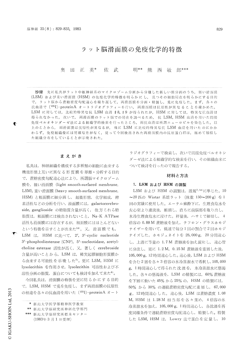
抄録 先に私共がラット中枢神経系のマイクロゾーム分画から分離した新しい膜分画のうち,軽い滑面膜(LSM)および重い滑面膜(HSM)の免疫化学的特徴を明らかにし,且つその細胞局在を明らかにする目的で,ラット脳から庶糖密度勾配遠心を繰り返して,両滑面膜を分画・精製し,兎に免疫した。まず,各々の抗血清で〔125I〕—proteinAオートラジオグラフィーを行い,両滑面膜は抗原性が異なることを確かめた。LSMに対しては,比較的特異な抗LSM 血清#8,#9が得られたが, HSMに対しては,特異な抗血清は得られなかった。次いで,両滑面膜のラット脳での局在を調べるため,抗LSM,HSM血清を用いた間接免疫ペルオキシダーゼ法による組織学的検索を行ったところ,両抗血清は所謂ニューロピルを染色した。以上のことから,両滑面膜は抗原性が異なるが,殊にLSMに比較的特異な抗LSM 血清を用いたのにかかわらず,免疫組織像には明確な差がなく,従って今回検出された両滑面膜内の抗原蛋白群は,極めて類似した組織分布をしていることが示唆された。
The microsomal fraction of the rat brain had been subfractionated by a sucrose density gradientcentrifugation into three distinct membrane frac-tions, namely "light smooth-surfaced membrane" LSM, "heavy smooth-surfaced membrane"HSM, and "rough-surfaced membrane". They were dif-ferent in their chemical compositions and enzym-atic activities as well as their buoyant densities.
In the present experiment antisera to purified LSM and FISM were raised in rabbits to charac-terize the two membranes immunochemically and immunohistochemically. Antigenicity of these mem-branes was studied by using 〔125I〕-protein A autoradiography and immunohistochemical locali-zation of the antigens in the rat brain was reveal-ed by the indirect immunoperoxidase technique. By 〔125I〕-protein A autoradiography (Fig. 1), dif-ferences in the antigenicity between LSM and HSM were clearly shown. Two out of nine anti-sera to LSM were relatively specific to LSM ; anti-LSM # 8 serum detected a protein band of Mr 53, 000 which is relatively specific to LSM (Fig. 1 B-a) and anti-LSM # 9 serum reacted with three proteins specific to LSM (Mr 53, 000, Mr 46, 000, and Mr 27, 000) (Fig. 1 C-a). However, anti-HSM sera did not discriminate between LSM and HSM antigens (Fig. 1 D & E) and non-immune serum did not detect any antigen at all (Fig. 1 F). Indi-rect immunoperoxidase pattern by anti-LSM # 8 revealed heavy staining of the neuropil in the cortices of cerebrum, hippccampus and cerebellum (Fig. 2 A, B & C). The perivascular structures identical to glial end-feet were also stained but the cell bodies and dendritic trunks of the neurons were not stained. The margin of some neurogliain the white matter was also stained (Fig. 2 D). Almost the same patterns were obtained by anti-LSM # 9 serum. The patterns by anti-HSM # 2 and # 3 sera seemed identical to those by anti-LSM sera (Fig. 3 A, B, C & D). In addition, the neuronal cell bodies bulk-isolated from the rat ce-rebral cortex were heavily stained by both anti-LSM and anti-HSM sera (Fig. 4 A, B & C). In complement fixation test the titer of antigen obtain-ed from the particulate fraction was higher than that from the soluble fraction of rat brains. So, the immunostaining of the neuropil may be attri-buted mainly to antigens of neuronal and glial membranes.
In summary, as revealed by immunoautoradio-graphy by anti-LSM # 8 and # 9 sera the antigeni-city of LSM proteins was different from that of HSM proteins. However, immunohistological pat-terns by these antisera seemed almost identical with those by anti-HSM sera. Following explana-tions can be made : ( 1 ) LSM and HSM distribute rather evenly and closely each other in the plasma membranes of neuropil including glial and neu-ronal membranes or ( 2 ) non-proteinous antigens common to LSM and HSM or high molecular we-ight proteins which reacted weakly to anti-LSM and common to LSM and HSM were responsible for the similarity in the immunohistochemical pat-terns between LSM and HSM. Future experiments with monoclonal antibody specific to LSM will unequivocally determine the localization of LSM in the nervous tissue.

Copyright © 1983, Igaku-Shoin Ltd. All rights reserved.


