Japanese
English
- 有料閲覧
- Abstract 文献概要
- 1ページ目 Look Inside
はじめに
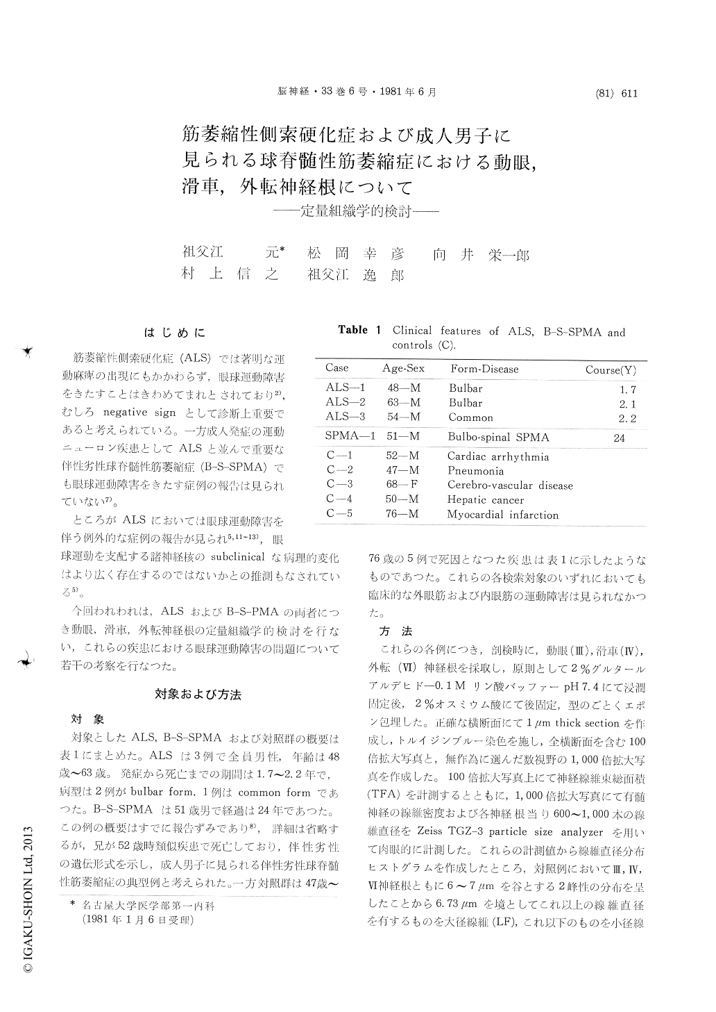
筋萎縮性側索硬化症(ALS)では著明な運動麻痺の出現にもかかわらず,眼球運動障害をきたすことはきわめてまれとされており2),むしろnegative signとして診断上重要であると考えられている。一方成人発症の運動ニューロン疾患としてALSと並んで重要な伴性劣性球脊髄性筋萎縮症(B-S—SPMA)でも眼球運動障害をきたす症例の報告は見られていない7)。
ところがALSにおいては眼球運動障害を伴う例外的な症例の報告が見られ5,11〜13),眼球運動を支配する諸神経核のsubclinicalな病理的変化はより広く存在するのではないかとの推測もなされている5)。
Morphometric quantification was performed on oculomotor, trochlear and abducent nerve roots from the cases of amyotrophic lateral sclerosis and X-linked recessive bulbo-spinal muscular atrophy. Three ALS cases were consisted of two bulbar forms and one common form aged 48 to 63 years old, whose clinical coures was 1.7 to 2.2 years (Table 1). The case of bulbo-spinal muscular atrophy aged 54 years old was suffered from muscular weakness and atrophy in bulbar musculature and extremities for more than 24 years. One of his siblings was died from similar disease at the age of 52 years. This case was considered to be a typical case of X-linked recessive bulbo-spinal muscular atrophy (B-S-SPM) based on its heredity form, clinical characteristics and anatomical findings that involved lower motor neurons. Five control cases of comparable age, free from neurological symptoms were analyzed in the same manner.
The results of morphometry on control casas revealed bimodal pattern in their fiber size frequency on these three nerve roots, as previously reported by other authors. Therefore, we could devide myelinated fibers into two major groups by fiber diameters at the division point of 6.73μm.
Analysis on the numbers of total myelinated fibers, large myelinated fibers and small myelinated fibers of these three nerve roots did not show any significant difference between ALS, B-S-SPMA and controls. Fiber size histograms on these roots from ALS, B-S-SPMA and contols revealed no significant difference in their bimodal patterns.
These results indicated that large myelinated fibers which considered to be α-motoneuron fibers were well preserved in these nerve roots, alhtough they were remarkably decreased in the spinal nerve roots of ALS and B-S-SPMA.
This difference on the root pathology between ventral spinal roots and oculomotor, trochler and abducent nerve roots will be comparable to the clinical features that external ocular muscles are selectively preserved in these disease, even in the advanced stage. These findings might also provide the clue to reveal the pathophysiology in α-motoneurons of ALS and B-S-SPMA.

Copyright © 1981, Igaku-Shoin Ltd. All rights reserved.


