Japanese
English
- 有料閲覧
- Abstract 文献概要
- 1ページ目 Look Inside
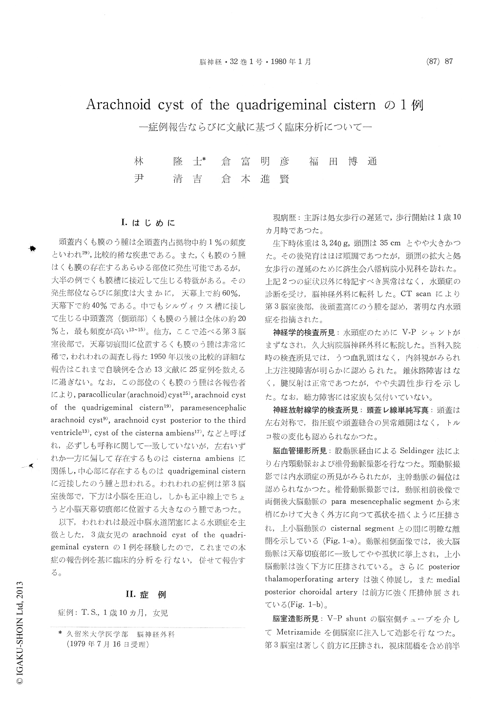
I.はじめに
頭蓋内くも膜のう腫は全頭蓋内占拠物中約1%の頻度といわれ29),比較的稀な疾患である。また,くも膜のう腫はくも膜の存在するあらゆる部位に発生可能であるが,大半の例でくも膜槽に接近して生じる特徴がある。その発生部位ならびに頻度は大まかに,天幕上で約60%,天幕下で約40%である。中でもシルヴィウス糟に接して生じる中頭蓋窩(側頭部)くも膜のう腫は全体の約20%と,最も頻度が高い13〜15)。他方,ここで述べる第3脳室後部で,天幕切痕間に位置するくも膜のう腫は非常に稀で,われわれの調査し得た1950年以後の比較的詳細な報告はこれまで自験例を含め13文献に25症例を数えるに過ぎない。なお,この部位のくも膜のう腫は各報告者により,paracollicular (arachnoid) cyst25),arachnoid cystof the quadrigeminal cistern19),paramesencephalicarachnoid cyst9),arachnoid cyst posterior to the thirdventricle13),cyst of the cisterna ambiens17),などと呼ばれ,必ずしも呼称に関して一致していないが,左右いずれか一方に偏して存在するものはcisterna ambiensに関係し,中心部に存在するものはquadrigeminal cisternに近接したのう腫と思われる。われわれの症例は第3脳室後部で,下方は小脳を圧迫し,しかも正中線上でちょうど小脳天幕切痕部に位置する大きなのう腫であつた。
Intracraial arachnoid cyst may occur in every site where the arachnoid exists, and it can be seen in the middle fossa, convexity, interhemisphere, parasellar, paracollicular (incisural), cerebellopontine angle and retro cerebellar regions. There are two theoretical opinions about its pathogenesis. One is that an abnormal dilatation of the subarachnoid space coinciding with the defective space due to cerebal malformation occurs, which forms a cyst after its loculation (subarachnoid cyst). The other is that an intraarachnoid cyst is formed due to anomaly in systemic genetic process of the subara-chnoid space.
In this paper, the authers described a case report of an arachnoid cyst of the quadrigeminal cistern and clinical analysis of this disease based on twenty-five published cases since 1941.
As a result, the age of onset ranged between three weeks old and thirty-seven years old. Among twenty-five cases, twenty-two cases (88%) were under fifteen years old, and suckling cases could be seen in ten cases (40%). On clinical symptoms, intracranial hypertension syndrome such as enla-rged head, sunset eyes, headache, papilledema etc. because of hydrocephalus due to obstruction of the aqueduct by the cyst could be seen in all cases. Vertebral arteriogram revealed an increases distance between the distal segment of the posterior cerebral and superior cerebellar arteries. Moreover, forward and downward displacement of the cho-roidal loop of the posterior inferior cerebellar artery and the basilar artery being closer applied the clivus.
On CT cisternogram of our case, it was identified the communication between the cyst and its adjacent subarachnoid space, namely the finding showed filling into the cyst and delayed clearance from it.
Their clinical features and operative considera-tions were discussed.

Copyright © 1980, Igaku-Shoin Ltd. All rights reserved.


