Japanese
English
- 有料閲覧
- Abstract 文献概要
- 1ページ目 Look Inside
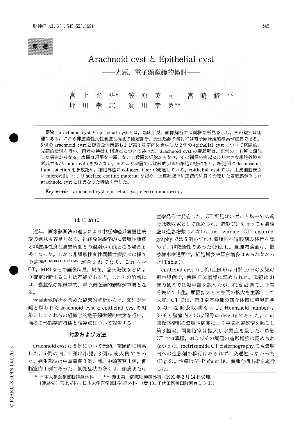
arachnoid cystとepithelial cystとは,臨床所見,画像解析では同様な所見を示し,その鑑別は困難である。これら非腫瘍性良性嚢腫性病変の確定診断,発生起源の検討には電子顕微鏡的検索が重要である。5例のarachnoid cystと傍四丘体槽部および第4脳室内に発生した2例のepithelial cystについて電顕的,光顕的検索を行い,両者の特徴と相違点について述べた。arachnoid cystの嚢腫壁は,正常のくも膜に類似した構造からなる。表層は扁平な一層,ないし数層の細胞からなり,その細長い突起により大きな細胞外腔を形成するが,microvilliを持たない。それより深層では比較的明るい細胞が密にあり,細胞間にdesmosome,tight junctionを多数認め,細胞外腔にcollagen fiberが発達している。epithelial cystでは,上皮細胞表面にmicrovilli,およびsurface coating materialを認め,上皮細胞下に連続的に良く発達した基底膜がみられarachnoid cystとは異なった特徴を示した。
There may be several kinds of pathlological con-ditions in the cystic lesion which are clinically diagnosed as benign intracranial cysts on CT scan.
Light and electron microscopic studies on cyst walls were important in the differential diagnosis of benign intracranial cysts. We have studied 5 cases of intracranial arachnoid cysts and two epithelial cysts using the light and electron microscopy.
Five cases of intracranial arachnoid cysts includ-ed two children and three adults (three females and two males) . Three cases of them were localized in the middle cranial fossa, one case in the anterior and middle cranial fossa and one case in the lateral ventricle, giving headache and convulsion as the initial complaints. As for the epithelial cysts, one was localized at the paracollicular area complainig enlarged head and swollen anterior fontanelle and the other of four years was located in the fourth ventricle with headache and ataxic gait. On CT all of them demonstrated diffuse low density areas in both the arachnoid and the epithelial cysts without communicating findings between the cystic cavities and subarachnoid space on metrizamide CT cister-nography.
The arachnoid cyst walls were basically similar in structure to the normal arachnoid membrane and composed of elongated epithelial cells like the ara-chnoid cell and the connective tissues with lamellar collagen fiber bundles. However, 3 of the 5 caseshad only fibrous tissues without epithelial cells. The inner sheath of the arachnoid cyst walls was com-posed of one or several layers of the arachnoid cells with flattened and relatively electron-dense cyto-plasm on electron micrograph. They had a lot of elongated process and were tangled with each other, making large extracellular spaces between them. Below the electron dense arachnoid cells, compact packed cells with interdigitation partly demonstrat-ed intercellular contacts such as numerous des-mosomes and tight junctions. In those intercellular spaces collagen fibers and microfibrils were obser-ved. The cells contained abundant cytoplasmic microfibrils and numerous organelles. They were seperated from numerous collagen fibers and fibro-blasts by non continuous basal lamina under the epithelial cells.
Epithelial cyst wall had a layer of cuboidal or columnal epithelium in the inner layer of cyst wall. Those epithelial cells demonstrated granules having positive in PAS and mucicarmine stain in their cytoplasm. On electron microscopical study epith-elial cells revealed a lot of microvilli and coating materials on the surface of them without cilia. The basement membranes were well developed under the epithelial cells separated from the connective tissues. In the intercellular clefts of the epithelial cells tight junctions and interdigitations were recog-nized. There were abundant ribosome, many mito-chondria, and dense granular materials in the cyto-plasm of the epithelial cells.
According to the electron microscopical study on the fine structure of the arachnoid cyst walls and the epithelial cyst walls the chracteristics and the differences in both of them were reported in this paper.

Copyright © 1991, Igaku-Shoin Ltd. All rights reserved.


