Japanese
English
- 有料閲覧
- Abstract 文献概要
- 1ページ目 Look Inside
I.はじめに
頭蓋頸椎移行部に発生したくも膜嚢胞は,cranio—spinal arachnoid cystと呼ばれ,極めてまれである.われわれは小脳半球内から上部頸椎椎管内に至る同症例を経験し,治療し得たので,文献的考察を加えて報告する.
We encountered a rare case of craniospinal arachnoid cyst. A 54-year-old woman was admitted to our clinic for headache, posterior nuchal pain, and vomiting, which had started one month before.
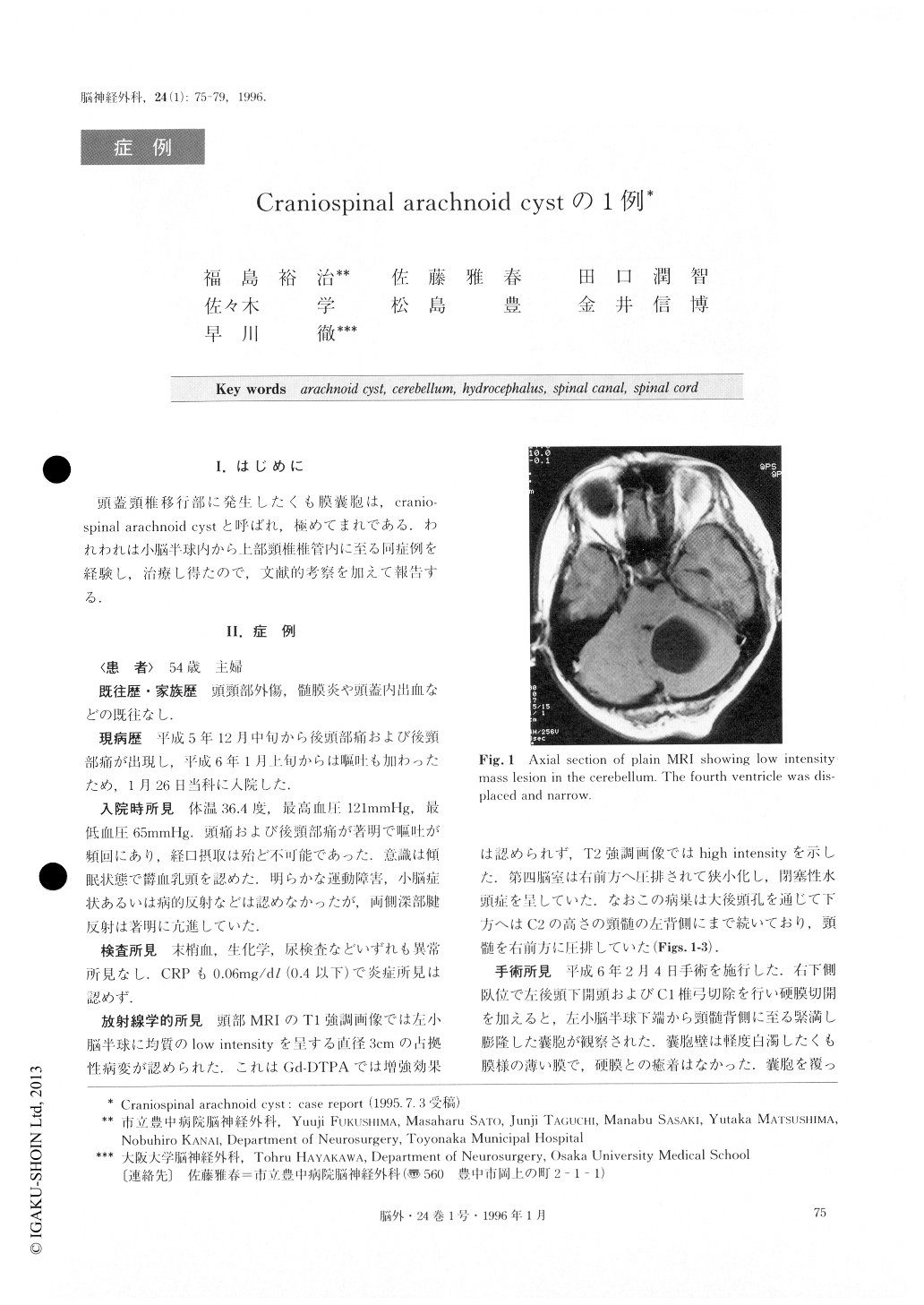
On admission she was drowsy, and neurological ex-amination revealed papilledema and prompt deep ten-don reflexes bilaterally. T1 weighted images of MRI re-vealed a homogeneous, round, low density mass in the left cerebellar hemisphere which extended clown on the dorsal side of the spinal cord to the level of C2. It was high intensity on T2 weighted images and was not en-hanced by Gd-DTPA. The fourth ventricle was com-pressed and obstructed and hydrocephalus was observed.
An operation was performed via a left suboccipital craniectomy with C1 laminectomy. A large cyst was lo-cated in the left cerebellar hemisphere, and it extended into the spinal canal clown to the C2 level where the cyst compressed the spinal cord right-anteriorly. The content of the cyst was xanthochromic fluid. After the removal of as much of the cyst as possible, the flat-tened tonsil was seen just dorsal to the medulla. The cyst extended into the cerebellar hemisphere via the widened fissura secunda. The arachnoid of the cisterna magna was incised and communication between the cis-terna magna and the cyst was made. The cyst was markedly reduced on the following day, and dis-appeared on the 50th postoperative day. The patient was discharged without neurological deficit. Pathologic-al diagnosis of the cyst wall in the cerebellum was thatthe molecular layer of the cerebellar cortex was co-vered by arachnoid membrane, which indicated arach-noid cyst.
Five such cases have been reported apart from this one. The therapy is the extirpation of the cyst wall and making communication between the arachnoid cyst and the subarachnoid space. The etiology of craniospinal arachnoid cyst is not well known but postoperative prognosis is good.

Copyright © 1996, Igaku-Shoin Ltd. All rights reserved.


