Japanese
English
- 有料閲覧
- Abstract 文献概要
- 1ページ目 Look Inside
I.はじめに
心臓血管性求心刺激は,血液量に関する低圧系と,血圧の維持調節機構に関する高圧系に分けられる8,12)。前者からはstretch receptor後者からはbaroreceptorによつて,主として迷走神経を介して,脳幹のvasomotorcenterおよびその周辺に対して,それらの情報が送られていることが報告されている5,7,8,11〜16,19)。しかし,心血管系からの大脳皮質への投射に関する研究はまだ少なく,Bailey1),Scheibel17)およびKornらが9),前頭葉の心窩部にそれからの投射を報告しているにすぎない。
本実験で観察した心臓血管性皮質電位に関しては,Callawayらが3),心臓サイクルと反応時間の研究のなかで,脳波を加算平均してそれを大きな徐波成分として記載しているが,詳しい解析は行なわれていない。
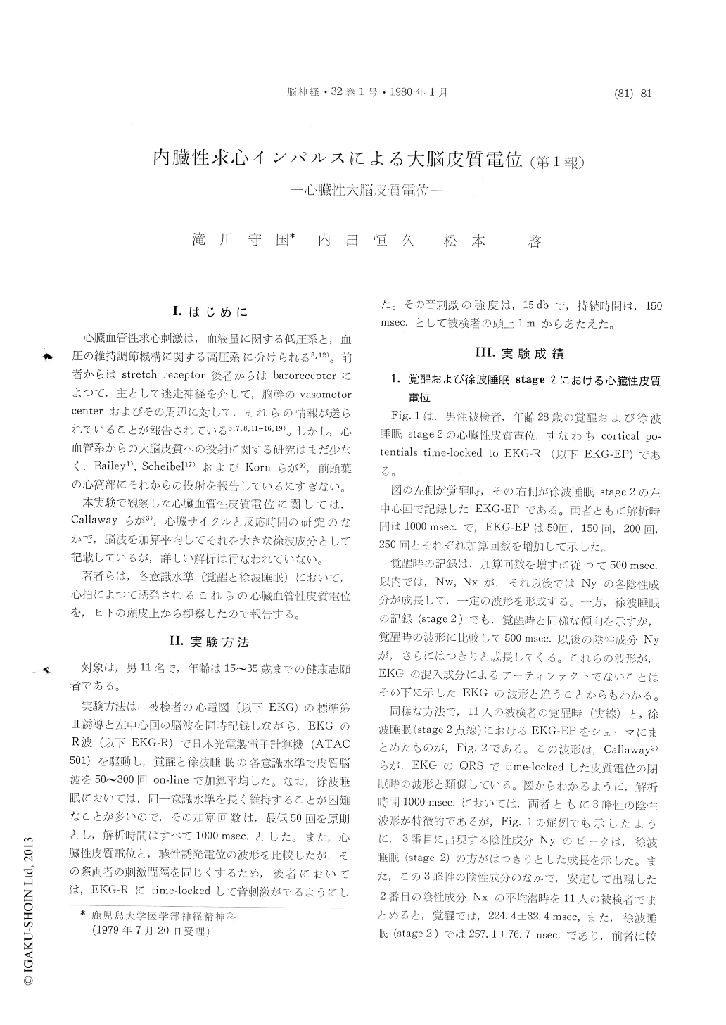
The purpose of this paper was to investigate whether cardiovascular afferent impulses could berecorded on a human scalp as computer-averaged cortical potentials time-locked to EKG-R waves.
The results obtained were as follows.
1) Cortical potentials time-locked to EKG-R waves (EKG-EP) could be recorded on scalps of eleven healthy volunteers and within its pattern, triple negative large peaks could be seen during both wakefulness and slow wave sleep (stage 2).
2) Within triple negative large peaks, the 2nd negative peak were most stable and its averaged latencies were 224.1±32.4 msec. during wakefulness and 257.1±76.7 msec. during slow wave sleep (stage 2) and the 3rd negative peak had been found more frequently in the slow wave sleep than in the wakefulness.
3) EKG-EPs were analogous with those of auditory evoked potentials time-locked to EKG-R waves (EKG-AEP) if both were recorded in the same concious level.
4) Both latencies of the 2nd negative peak of the EKG-EPs and that of the largest negative peak of the EKG-AEPs were consistent each other with high ratio resulting r=0.81 (P<0.01) in the wakefulness and r=0.966 (P<0.001) in the slow wave sleep (stage 2) respectively.

Copyright © 1980, Igaku-Shoin Ltd. All rights reserved.


