Japanese
English
症例
Suprasellar arachnoid cystの1例
Suprasellar Arachnoid Cyst:Report of a case
高橋 徳
1
,
川合 省三
1
,
上農 哲朗
1
,
平松 謙一郎
1
,
前川 基継
1
,
湯浅 隆史
1
,
宮元 直数
1
,
服部 裕
1
Toku TAKAHASHI
1
,
Shozo KAWAI
1
,
Tetsuro KAMINOH
1
,
Ken-ichiro HIRAMATSU
1
,
Mototsugu MAEKAWA
1
,
Takashi YUASA
1
,
Naokazu MIYAMOTO
1
,
Yutaka HATTORI
1
1大阪府立病院脳神経外科
1Department of Neurosurgery, Osaka Prefectural Hospital
キーワード:
Suprasellar lesion
,
Arachnoid cyst
,
Hydrocephalus
,
CT
Keyword:
Suprasellar lesion
,
Arachnoid cyst
,
Hydrocephalus
,
CT
pp.435-441
発行日 1982年4月10日
Published Date 1982/4/10
DOI https://doi.org/10.11477/mf.1436201501
- 有料閲覧
- Abstract 文献概要
- 1ページ目 Look Inside
I.はじめに
くも膜嚢腫はその特徴的な所見18,20),のためにCTにより比較的容易に診断しうることから経験される機会が増加してきているが,髄液腟との交通性の有無や手術法の検討など幾つかの問題点をもっている23).著者らは本邦においては報告の少ない鞍上部くも膜嚢腫の1例を経験したので,主として手術方法を中心に若干の文献的考察を併せて報告する.
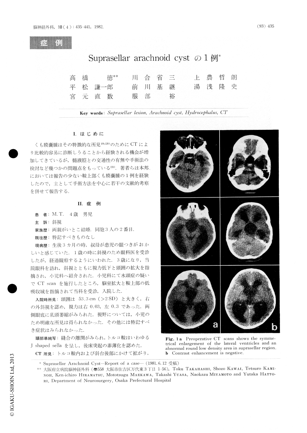
A 4-year-old boy with suprasellar arachnoid cyst wasreported. At the age of 3-month-old his aunt was awareof his squint. During the observation by ophthalmologistsfrom the age of ly. to 3y., enlargement of the head and im-pairment of the visual acuity were manifested. Cranial CTscan revealed the enlargement of the ventricular systemand a round low density area located superior to the sella.Absorption coefficient of the lesion was similar to that ofthe cerebrospinal fluid. No abnormal contrast enhancementwas seen.

Copyright © 1982, Igaku-Shoin Ltd. All rights reserved.


