Japanese
English
- 有料閲覧
- Abstract 文献概要
- 1ページ目 Look Inside
はじめに
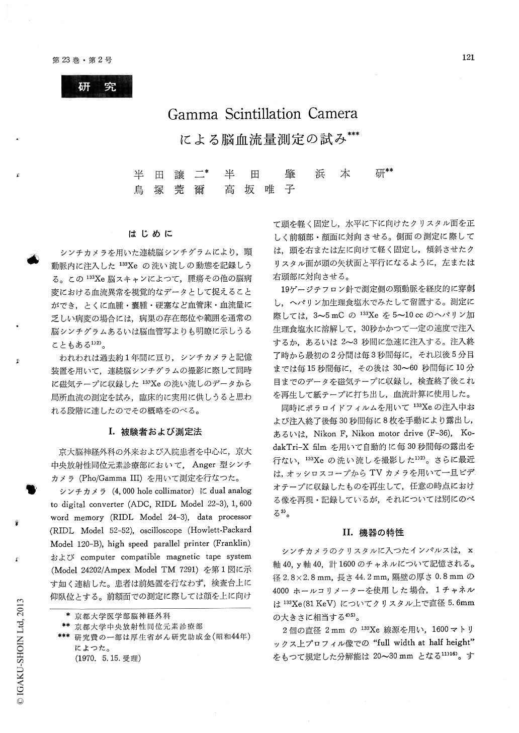
シンチカメラを用いた連続脳シンチグラムにより,頸動脈内に注入した133Xeの洗い流しの動態を記録しうる。この133Xe脳スキャンによつて,腫瘍その他の脳病変における血流異常を視覚的なデータとして捉えることができ,とくに血腫・嚢腫・硬塞など血管床・血流量に乏しい病変の場合には,病巣の存在部位や範囲を通常の脳シンチグラムあるいは脳血管写よりも明瞭に示しうることもある1)2)。
われわれは過去約1年間に亘り,シンチカメラと記憶装置を用いて,連続脳シンチグラムの撮影に際して同時に磁気テープに収録した133Xeの洗い流しのデータから局所血流の測定を試み,臨床的に実用に供しうると思われる段階に達したのでその概略をのべる。
1. Quantitative measurements of regional cerebral blood flow were performed by the use of 133Xe and the gamma scintillation camera (Pho/Gamma III) equipped with 1600 channel memory, data processor, computer compatible magnetic tape system and high speed printer.
2. After intracarotid injection of 133xe in saline, the activity of the brain was sampled on 1600 matrices and recorded for 10 minutes. Washout data were analysed and the flow index (ratio be-tween the total counts on a given matrix for initial 15 seconds and total counts on the same matrix for 10 minutes) was calculated for each matrix and mapped out. The pattern of distribution of flow indices compared favorably with the results obtained by several other methods of blood flow measurements.
3. For the purpose of compartmental analysis of the washout curve, 4, 6 or 9 matrices was combined and used for 1 ROI (region of interest). The results obtained were correlated with localisation of the tumor or other intracranial pathology. Generalizeddecrease in cerebral blood flow due to increased in-tracranial pressure, abnormal tumor flow pattern with or without shunt peak, and perifocal zone ofischemia were well demonstrated.

Copyright © 1971, Igaku-Shoin Ltd. All rights reserved.


