Japanese
English
- 有料閲覧
- Abstract 文献概要
- 1ページ目 Look Inside
(1)scintillation camera (PHO/GAMMA III)と短半減期RI(99mTc)を用いると,短時間に,任意の方向から脳scintiphotoを撮影でき,被検者の曝射,時間的制約を最少限におさえられる。さらに1回のRI投与後くりかえしscanを行なうことにより,RIとりこみ像の動的な変化を知ることができる。
(2)さらに1600 channel memory scopeおよびprint-erの使用によりよわいとりこみの検出も可能であるほか,RIとりこみの時間的変化を数量的に示すことができる。この方法を用いて.各種病巣の99mTcとりこみ比の時間的変化をみると,(1)漸減型(動静脈奇形,血管腫,meningiornaなど),(2)漸増型(脳軟化,良性gliomaなど),(3)漸増後減少型(glioblastoma, sarcoma, car-cinomaの多くのものなど)および,(4)coolな病巣(血腫など)に分けることができた。
(3)133Xe, 131I-MAAの頸動脈内注入後の脳scntiscan-ningの成績および血管写所見から第1型のとりこみは主として病巣内血管床・血流量の増大によつて規定される。第2,4型の病巣は常に血管床・血流量に乏しい。第3型はこの両者の混合ないし中間型と考えられる。
(4)脳scintiscanningにおける動的観察の意義について述べた。
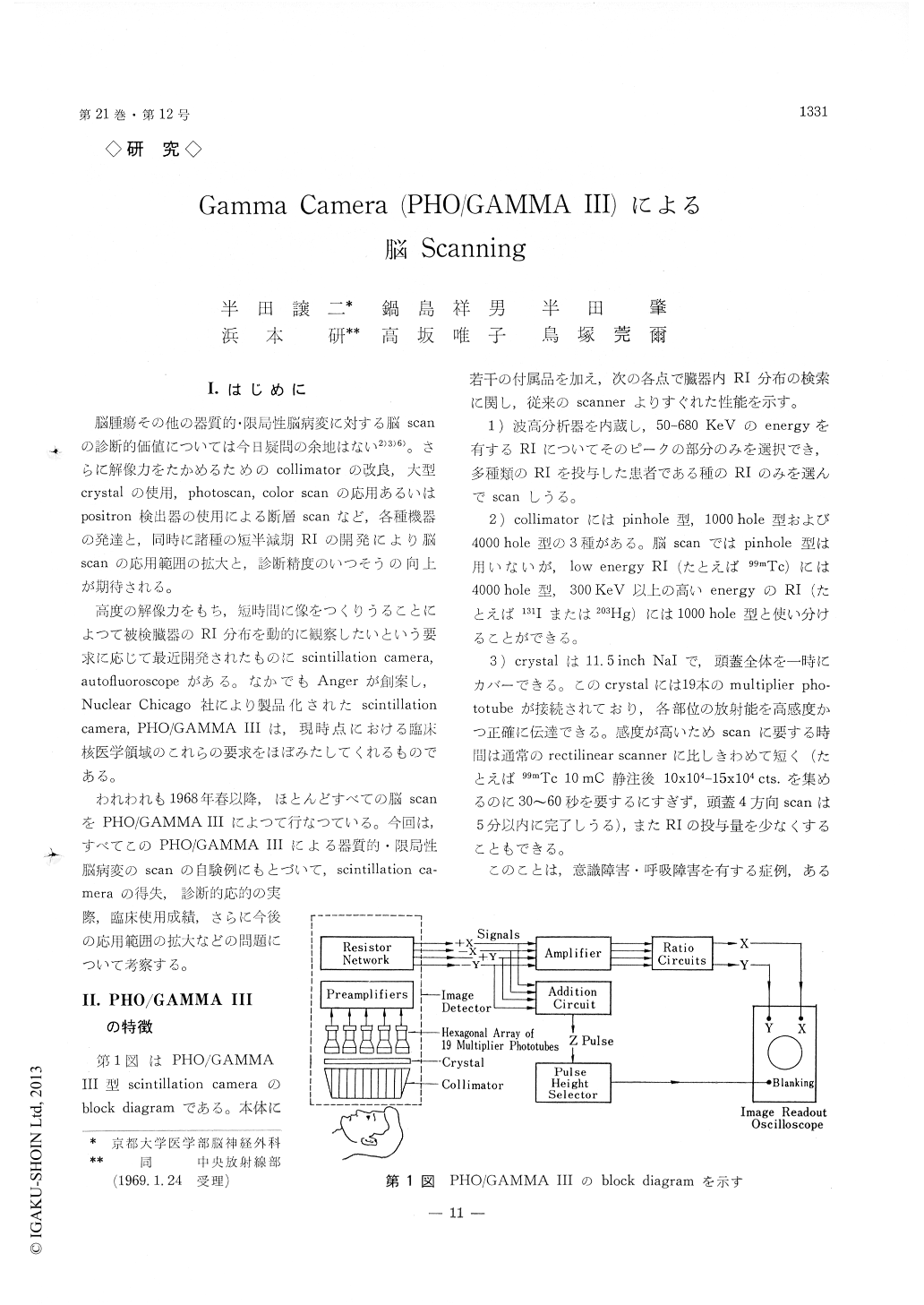
The sensitivity of the scintillation camera with multichannel collimators is appreciably higher than focused-collimator scanners. The resulting greater speed of scanning is a definite advantage for neu-rosurgical cases. A number of different views can be taken as desired. The examination can he com-pleted in a short time, this provides us with the possibility of serial scanning following a single dose of tracer, and a dynamic analysis of the uptake pattern is possible.
By use a memory scope and 1600 channel multi-parameter analyzer, the uptake ratio can be calculated serially and correlated with the interval between the administration of a tracer and the examination. The time course of the uptake ratio seems to be divided into four main categories. In type 1, the uptake ratio reaches the maximum immediately or shortly after the intravenous injection of 99mTc, then it decreases rapidly. In type 2, the uptake ratio shows a gradual increase during the examination period up to 2 hours. In type 3, the uptake ratio shows an initial rise, reaches the maximum 30 to -15 minutes after theintravenous administration of 99mTc, then it decreases again. In the type 4, the uptake ratio remains below 1, namely the focus is cool.
Meningiomas, arteriovenous malformations and one case of cavernous angioma belonged to type 1, astro-cytomas, oligodendrogliomas and cerebral infarctions to type 2, glioblastomas, sarcomas and most of the metastatic carcinomas to type 3. Uptake pattern of type 4 was seen in intracerebral hematomas.
Serial brain scanning following intracarotid injec-tion of 133Xe or 131I-MAA permits the estimation of blood flow or vascular bed in the pathologic focus. It has been definitety shown that the high blood flow and rich vascularity of the focus are the main factors responsible for the uptake pattern of type 1. In contrast, in the lesion which showed the uptake course of types 2 and 4, the focus was pooly vascu-larized.
Copyright © 1969, Igaku-Shoin Ltd. All rights reserved.