Japanese
English
- 有料閲覧
- Abstract 文献概要
- 1ページ目 Look Inside
I.はじめに
Computed Tomography (CT)が開発され,その目覚しい発達,普及とともに神経放射線診断領域に大きな変化をもたらしつつある。著者らも1976年6月以来CT(CT−1000(EMI社))スキャナの使用の機会を得ているが,本装置の特徴として,ある断層面における組織のX線吸収値の分布を0.5%の精度9)で表示すること,この検査が安全かつ容易で患者のX線被爆量も少ないことなどがあげられる1,5,11)。また従来のX線フイルムでは最適条件であつても2〜4%のContrastがなければ検出できないこと15,22)に比較しても画期的な装置である。このようにCTにより従来のX線フイルムとはその質,量とも全く異つた画像情報が得られるようになつたが,この情報をいかに有効に活用するか,すなわち日常の診断においてその精度を高め,またいかに有効な治療に結びつけるかが今後の課題になつている。
従来の線診断では,まずX線フイルムからできるだけ多くの特徴の抽出,すなわち特徴抽出の過程,次にその特徴と臨床データ,我々のもつ解剖・病理的知識あるいは過去の経験などから総合的に判断し最終的な診断を行うという過程,すなわち認識の過程をとつてきた19)。CTの診断においても画像情報そのものは異つているが,診断の過程においては従来のX線診断と変わるところがない。
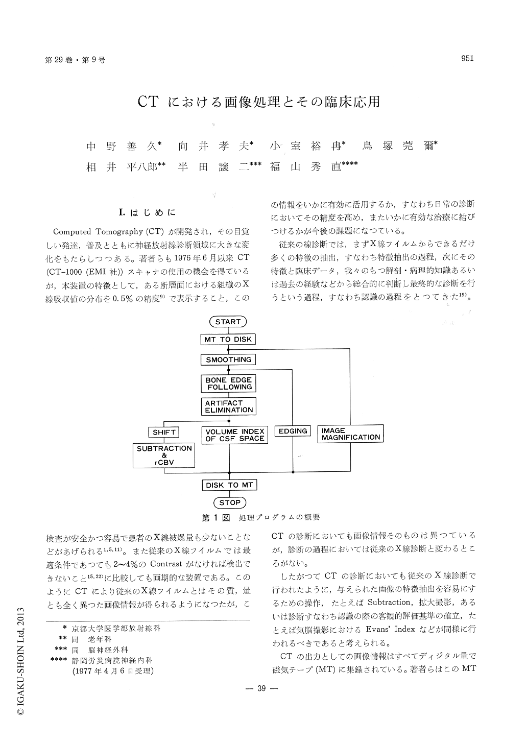
Using the digital data stored on the magnetictape of the EMI scanner system (EMI head scanner,model 1000), computer programs for "Smoothing","subtraction", "edging" and measurements of"regional cerebral blood volume (rCBV)" and"volume index of cerebrospinal fluid (CSF) space"have been developed. Processing was done by aData General, Eclipse S/200 or DEC, PDP 11/40computer and programs were written in FORTRAN4. Processed pictures were restored on the magnetictape and displayed on the ordinary display unit ofthe EMI scanner system.
Smoothing was performed by simply replacingthe CT number at a given picture element (pixel)by the average of 9 values over a 3×3 pixelscentered at that particular pixel. Weighted aver-aging or other nonlinear operations involving theaveraging were also performed. Without smoothing,a wide window setting on the display unit is neces-sary to reduce noise in the pictures, however, thistends to lessen the possibility of detecting the smalldifferences in attenuation. Digital smoothingreduced noise in the pictures by itself, so that therecognition of the large regions with slight differ-ences in attenuation was possible even with anarrow window setting.
Subtraction is useful to take full advantage ofcontrast enhancement technique. When subtractionwas performed without preparatory smoothing,however, this technique often fails to improve thedetectability of small changes in attenuation, sincethe noise in the original pictures is propagated.To suppress noise in the subtracted picture,smoothing of pre- and postcontrast pictures wasdone as an initial step and point-to-point subtractionwas computed after vertical and horizontal ad-justments for better superimposition of two pictures.Measurements of "γCBV" with CT was performedeasily by this subtraction method.
Edges or lines can be emphasized by performinga suitable differentiation operation. After smoothingwas done, edge features of the intracranial structuressuch as ventricles, cisterns or any lesions wereobtained by taking the derivatives in a gradientdirection. Edging proved useful, particularly indemonstrating periventricular edema in hydroce-phalus more clearly.
Percentage of the number of pixels with CTnumber of 10 or less to the number of whole pixelswithin the cranial cavity of the same slice wascomputed. This value denotes a two-dimensional(surface) index of the area of the CSF space tothat of the cranial cavity. Computing the datafrom four contiguous slices, a three-dimensionalvolume index of CSF space was obtained, whichproved useful in evaluating, for example, the degreeof cerebral atrophy or hydrocephalus, and thepatency of the CSF shunts.
Copyright © 1977, Igaku-Shoin Ltd. All rights reserved.