Japanese
English
- 有料閲覧
- Abstract 文献概要
- 1ページ目 Look Inside
まえがき
一側性眼球突出を訴える患者をみることはさほど稀ではなく,それは相異なる疾患の臨床症状の1つである8)9)19)。したがつて,その原疾患を正しく,早く診断することが適切な治療法を決定するために非常に必要である。
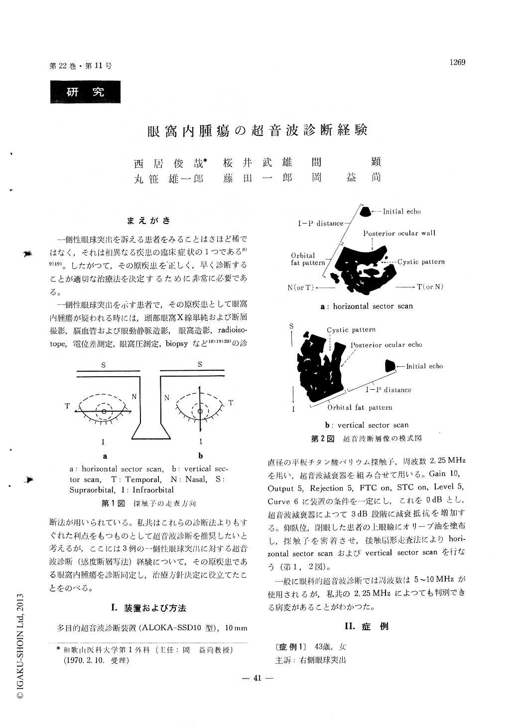
一側性眼球突出を示す患者で,その原疾患として眼窩内腫瘍が疑われる時には,頭部眼窩X線単純および断層撮影,脳血管および眼動静脈造影,眼窩造影,radioiso—tope,電位差測定,眼窩圧測定,biopsyなど18)19)20)の診断法が用いられている。私共はこれらの診断法よりもすぐれた利点をもつものとして超音波診断を推奨したいと考えるが,ここには3例の一側性眼球突出に対する超音波診断(感度断層写法)経験について,その原疾患である眼窩内腫瘍を診断同定し,治療方針決定に役立てたことをのべる。
Various diagnostic methods have been used in the diagnosis of unilateral exophthalmos suspected of the intraorbital tumors. One of these methods is the ultrasonic diagnostic method which is more suitable and simple than others. By this method we have successfully made diagnosis on 3 cases of the intra-orbital tumors, namely mixed tumor of the lacrimal gland, glioma of the optic nerve and lymphosarcoma including infant, which have developed unilateral exophthalmos. Other methods than ultrasonic diag-nosis have failed to make correct diagnosis.
For these cases we have used the ultrasonotomo-graphy with varying sensitivity by the contact scanning method. The ultrasonic pulses used are 2. 25 MHz in frequency and the intensity has been changed by 3 dB. The frequency of 2. 25 MHz seems to be slightly lower than usual in ophthalmologic use of 5 to 10 MHz.
Ultrasonic echo patterns have been found as the cystic pattern which is formed by the tumor itself, orbital fat pattern deformed by the tumor and I-P distance (length from starting point of inital echo to posterior ocular wall) which is shortened by the tumor.

Copyright © 1970, Igaku-Shoin Ltd. All rights reserved.


