Japanese
English
- 有料閲覧
- Abstract 文献概要
- 1ページ目 Look Inside
I.はじめに
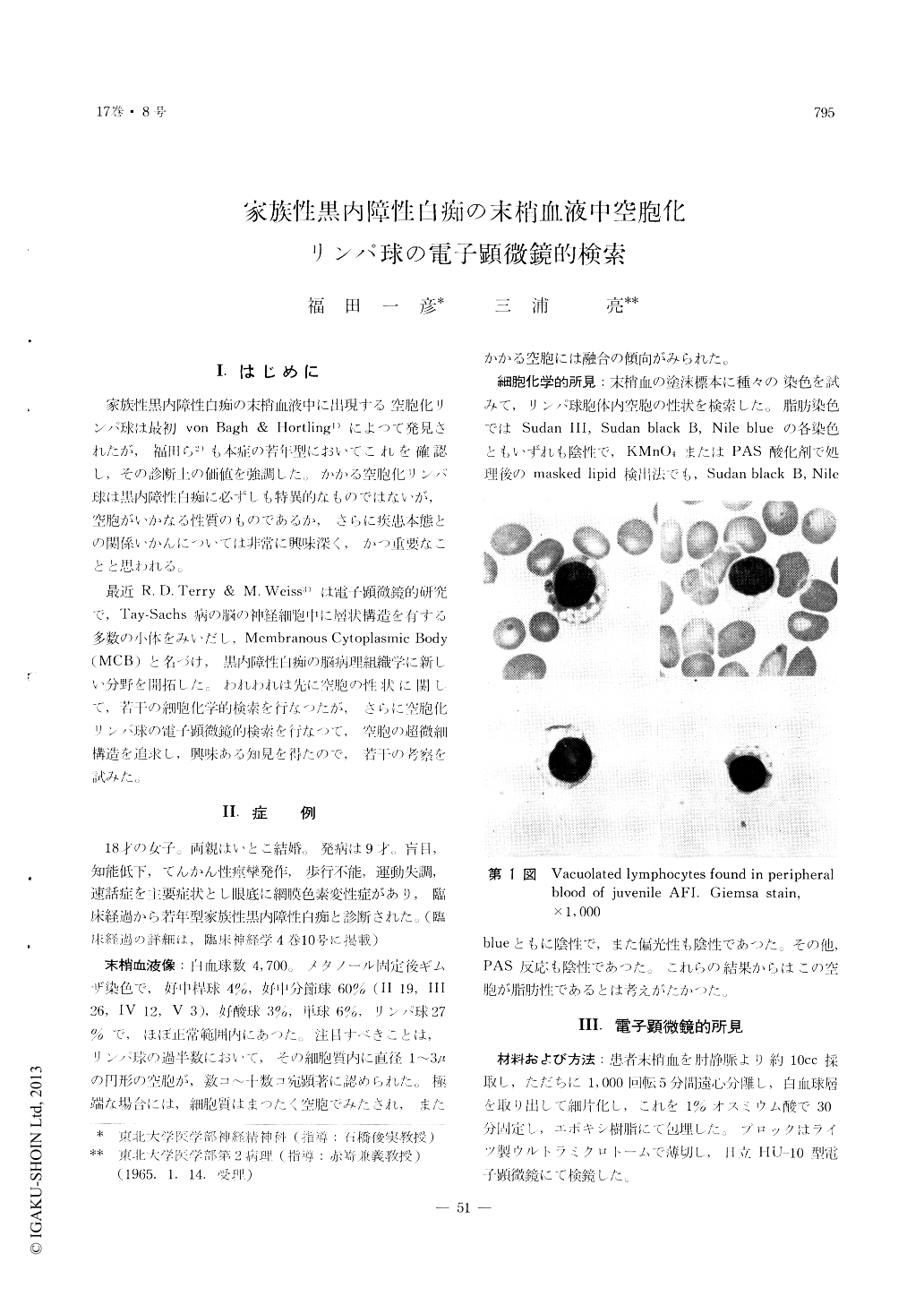
家族性黒内障性白痴の末梢血液中に出現する空胞化リンパ球は最初von Bagh & Hortling1)によつて発見されたが,福田ら2)も本症の苔年型においてこれを確認し,その診断上の価値を強調した。かかる空胞化リンパ球は黒内障性白痴に必ずしも特異的なものではないが,空胞がいかなる性質のものであるか,さらに疾患本態との関係いかんについては非常に興味深く,かつ重要なことと思われる。
最近R. D. Terry & M. Weiss3)は電子顕微鏡的研究で,Tay-Sachs病の脳の神経細胞中に層状構造を有する多数の小体をみいだし,Membranous Cytoplasmic Body (MCB)と名づけ,黒内障性白痴の脳病理組織学に新しい分野を開拓した。われわれは先に空胞の性状に関して,若干の細胞化学的検索を行なつたが,さらに空胞化リンパ球の電子顕微鏡的検索を行なつて,空胞の超微細構造を追求し,興味ある知見を得たので,若干の考察を試みた。
The cytochemical and electron microscopic ex-aminations were performed on the vacuolated lym-phocytes found in the peripheral blood of juvenile amaurotic family idiocy (AFI). Vacuoles were neither stained with Sudan III, Sudan black B and Nile blue, nor even with Sudan black B and Nile blue after the oxidation with KMnO4 or PAS oxidizer. These results seemed to lead us to the conception that the vacuoles would not contain lipid.
About a volume of 10cc of blood was taken from the cubital vein, which was centrifuged at 1,000 r. p. m. for five minutes. The layer of leucocytes were sliced and fixed in 1% osmi cacid for thirty minutes, then embedded in epon.
The examination of the ultrastructures of the cells which were identified as lymphocytes revealed very characteristic appearances. The abnormal corpuscles which are never present in normal lymphocytes were observed in the cells. The vacuoles found in Giemsa preparates may be coincident with these abnormal granules, which showed several appearances.
Round bodies of mitochondrial size with an outer smooth limiting membrane, surrounding an inner vacant zone partly filled with electron-dense, homo-geneous amorphous or parallel double membrane-like material, were most common. It was noteworthy that there were other unique bodies which were composed of two or three layers arranged concen-trically or had onion skin-like appearances. The amorphous material or parallel membranous body may be essentially the same as the amorphous form or parallel membranes which E. H. Mercer insisted upon on the evolution of intracellular phospholipid membrane system. The concentrically lamellated unusual structure may be substantially the same as that of one of the membranous cytoplasmic bodies (MCB) which R. D. Terry and M. Weiss found in the cytoplasm of the nerve cells of Tay-Sachs brain and which contain a large amount of lipid.
In conclusion, it seems likely that the vacuoles found in Giemsa preparates may contain lipid, con-sidering their fine structures. If it can be asserted that the MCB-like bodies are exactly the same as MCB found in the nerve cells of Tay-Sachs brain, the relation, or lack of it, between the vacuolated lymphocytes in the peripheral blood of juvenile AFI and the metabolic disturbance or the pathogenesis of the disease will be made clear.

Copyright © 1965, Igaku-Shoin Ltd. All rights reserved.


