Japanese
English
- 有料閲覧
- Abstract 文献概要
- 1ページ目 Look Inside
I.いとぐち
1913年Luckett1)によつて頭蓋底骨折部より脳底くも膜下腔を経て脳室内に到達した空気が,始めてレ線撮影により影像として確認されたが,頭蓋内空気像を臨床神経学的診断に応用したのはさらに5年後である。1918年,Dandy2)3)が腰椎穿刺により空気を人為的に脊髄くも膜下腔に注入し,これによつて脳室像を得ることに成功したのに始まり,ついで1920年にはBingel4)がDandyとは無関係に,彼と同様の方法を用いて脳室像のレ線による撮影に成功し,この方法をpneumoencephalographyと称して発表した。これらDandyおよびBingelの発見による気脳撮影法はその後,神経学の分野における診断および研究方法としてきわめて大なる貢献をなしてきており,血管撮影のめざましい普及をみた現在においても,なお,その価値を失なうものではなく広く行なわれてきている。
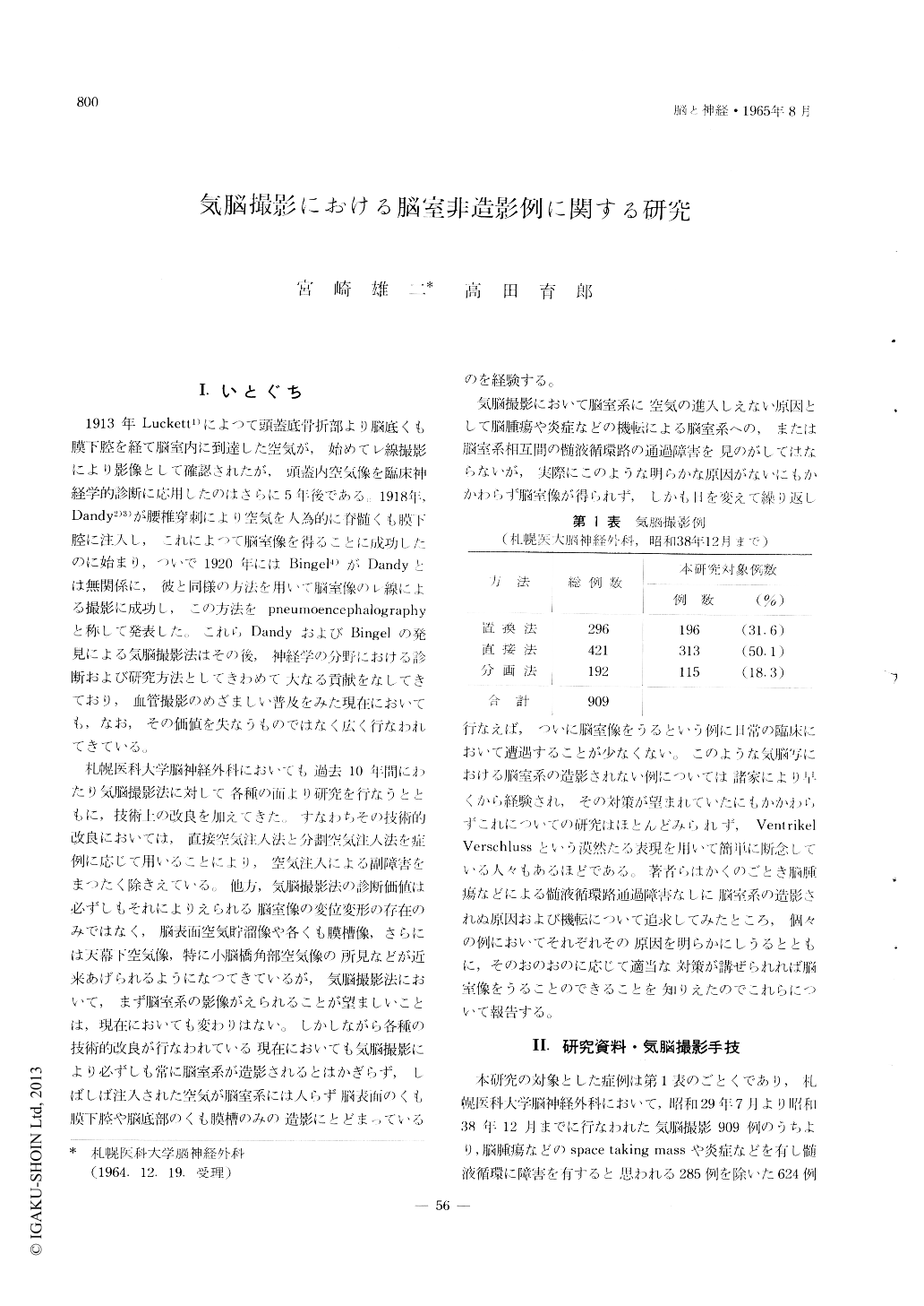
札幌医科大学脳神経外科においても過去10年間にわたり気脳撮影法に対して各種の面より研究を行なうとともに,技術上の改良を加えてきた。すなわちその技術的改良においては,直接空気注入法と分劃空気注入法を症例に応じて用いることにより,空気注入による副障害をまつたく除きえている。他方,気脳撮影法の診断価値は必ずしもそれによりえられる脳室像の変位変形の存在のみではなく,脳表面空気貯溜像や各くも膜槽像,さらには天幕下空気像,特に小脳橋角部空気像の所見などが近来あげられるようになつてきているが,気脳撮影法において,まず脳室系の影像がえられることが望ましいことは,現在においても変わりはない。しかしながら各種の技術的改良が行なわれている現在においても気脳撮影により必ずしも常に脳室系が造影されるとはかぎらず,しばしば注入された空気が脳室系には入らず脳表面のくも膜下腔や脳底部のくも膜槽のみの造影にとどまっているのを経験する。
Since Dandy's description in 1919 on the roent-genographical examination of the central nervous system by air injection through spinal subarachnoid space, pneumoencephalography has been used ex-tensively and this procedure has still diagnostic value in the field of neurology and neurosurgery in spite of present widespread appliation of cerebral angiography.
Many articles had published by various authors on the pneumoencephalography. The majority had dis-ccussed on development of the new techniques, complication and various pathological findings and very few papers on the cause of failure to fill the ventricular system in cases which has no pathological obstruction of cerebrospinal fluid pathway.
The purpose of this paper is to know the cause of unfilling of ventricular system in normal cases and to procedure to get ventricular shadow in such cases.
The 83 cases out of 909 cases which had done the pneumoencephalography in Neurosurgical department of Sapporo Medical College Hospital during past 10 years are submitted to this study as cases of non-filling of ventricular system. 83 cases were able to classify into six groups as following based on the intracranial location of air.
1st group: No air is visible intracranially.
2nd group: Localized air collection in Cisterna Magna.
3rd group: The air is visible mainly in basal ara-chnoidal cisterna.
4th group: The air is visible in dorsal arachnoidal cisterna of brain stem.
5th group: Combined and complicated cases of above 3rd and 4th groups.
6th egroup: Subdural air collection.
From the anatomical and hydrodynamic stand point the cause of unfilling of ventricular system was disccussed in each groups and procedure to get the ventricular filling in each group had also described.

Copyright © 1965, Igaku-Shoin Ltd. All rights reserved.


