Japanese
English
- 有料閲覧
- Abstract 文献概要
- 1ページ目 Look Inside
I.急性・亜急性頭蓋内血腫
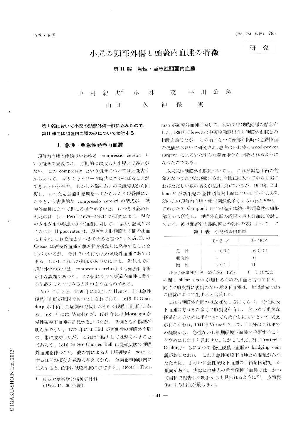
頭蓋内血腫の症候はいわゆるcompressio cerebriという概念で表現され,原則的には成人と小児とで違いがない。このcompressioという概念については大変古くからあつて,ギリシャ・ローマ時代にさかのぼることができるという26)76)。しかし外傷のあとの意識障害から回復し,いつたん意識明瞭期をへてからふたたび昏睡にいたるという古典的なcompressio cerebriの型式が,硬膜外血腫によつて起こる場合が多いと,はつきり認められたのは,J. L. Petit (1675-1750)の研究による。現今のさまざまの疾患や医学知識に関して,博学な記載をおこなつたHippocratesは,頭蓋骨と脳硬膜との間の出血にもふれ,これを除去すべきであると言つた。25A. D.のCelsusは硬膜外血腫が頭蓋骨骨折なしに発生することを述べているが,今日でいえば小児の硬膜外血腫にあてはまる、しかしこれらの知識があつたにせよ,近代までの頭部外傷の医学は,compresio cerebriよりも頭蓋骨骨折が主な課題であつた。この間にあつて頭蓋内血腫に関する記載をひろつてみると次のようなものがある。
Paréによると,1559年に死亡したHenry二世は急性硬膜下血腫が死因であつたとされており,1619年Glan—dorpが手術した症例の記載もおそらく硬膜下血腫である。1681年にはWepferが,1747年にはMorgagniが慢性硬膜下血腫の剖見例を述べたが,2例とも外傷歴が明らかでない。1772年にはHillが両側性の硬膜外血腫の手術に成功したが,これは当時としては驚くべきことであろう。1816年Sir Charles Bellは屍頭実験で硬膜外血腫を作つた6)。彼の言によると「脳硬膜をlooseにするほどの振動を屍頭に与えてから,色素を頸動脈内に注入すると,色素は硬膜外腔に貯溜する」。1828年Thor—manが硬膜外血腫に対して,初めて中硬膜動脈の結紮をした。1861年Hewettは中硬膜動脈出血と硬膜外血腫との相関を論じたが,この頃になつて頭部外傷時の意識障害の機構がおおいに研究され,患者はいわゆるwood-pecker surgeonによるいたずらな穿頭術から開放されるようになつたのである。
1) Four cases of acute and one case of subacute extradural hematoma were encountered in the neuro-surgical department of the University of Tokyo Hospital.
All cases were accompanied by skull fracture on the ipsilateral side of the hematoma, one of which was not demonstrated on the roentgen-film. Even if decerebrated state was evident on admission, early and proper operation could relieve the patient from the seriousness and grave condition, and a good prog-nosis for survival could be expected.
2) Six cases of acute and three cases of subacute subdural hematoma were encountered in the above mentioned Hospital.
Six patients out of a total of nine were below two years of age.
The severity of trauma was moderate or slight. The direction of blow was mainly to the occipital region of the head. Three cases were striking in the fact that although the trauma was slight, the patients lost their consciousness followed by con-vulsion, after a very short period of lucid interval. The clinical course afterwards very rapidly deteri-orated from bad to worst and immediate surgical intervention could save only one patient, leaving the remaining two in the hands of death.
The lucid interval followed by sudden and con-tinuous loss of consciousness with convulsion was the common typical feature of the early-stage sub-dural hematoma. The longest lucid interval in this series was seven days.
Retinal hemorrhage was found in six cases out of eight examined and therefore this phenomena could be considered as one of the important signs in the diagnosis of these subdural hematoma.
In six cases verified, the source of bleeding was the rupture of the bridging vein from the cortical surface to the superior longitudinal sinus or to the dural lacuna; and no cortical contusion or laceration was found.
The prognosis of survivors was excellent in gene-ral.

Copyright © 1965, Igaku-Shoin Ltd. All rights reserved.


