Japanese
English
- 有料閲覧
- Abstract 文献概要
- 1ページ目 Look Inside
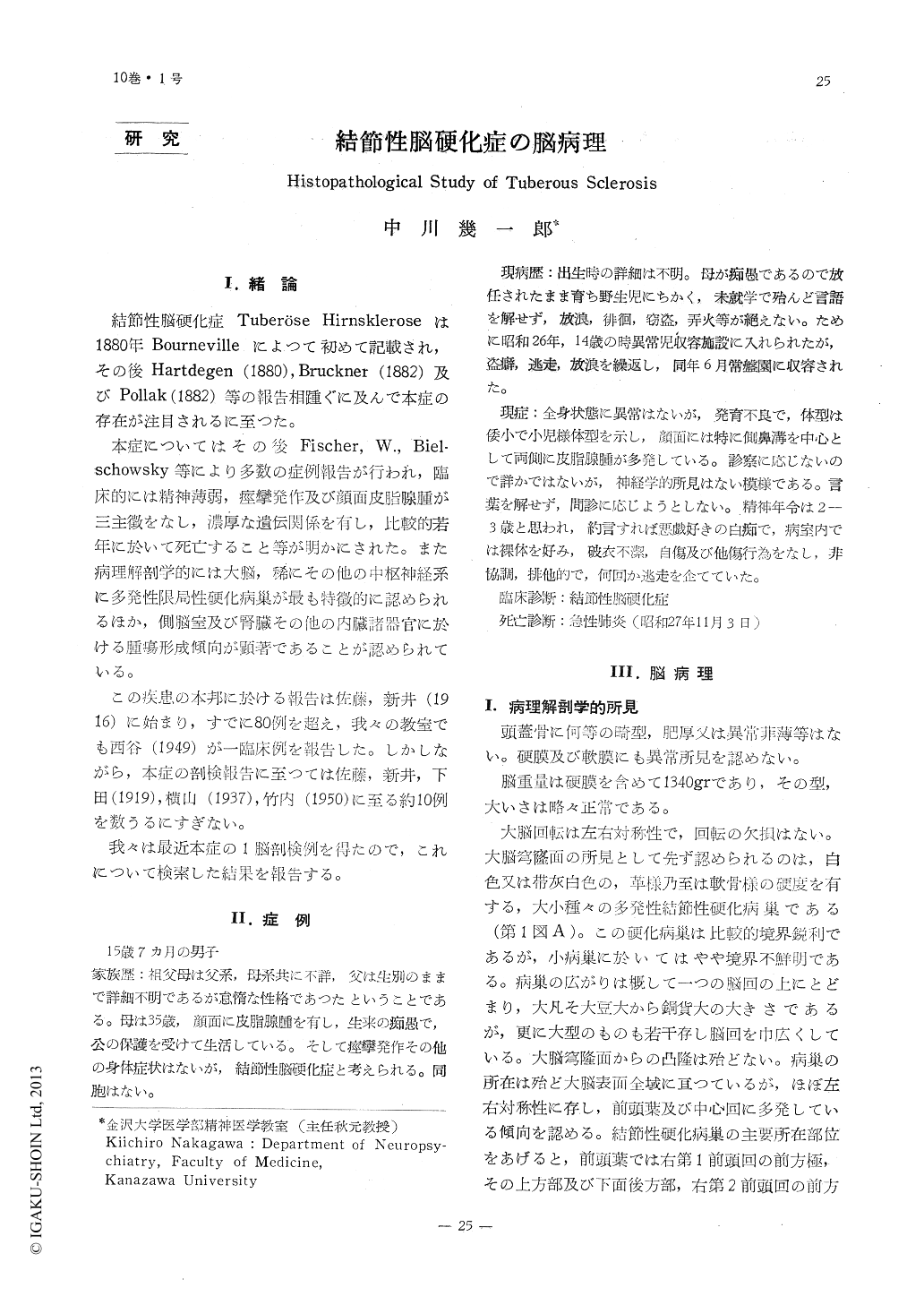
臨床上精神薄弱(白痴)と顔面皮脂腺腫を有した結節性脳硬化症の1脳を剖検し,次の所見をえた。
1.主なる病変は大脳の殆ど全域に存する硬化 病集と側脳室腫瘍である。
2.大脳硬化病巣は肉眼的に腫瘍像を呈するも の(第2型病巣Pellizi)はな・く,膠線維増生の 盛なグリオーゼのほか神経細胞の脱落,変性及 び異型巨大細胞の退行性変化の像を認める。
3.大脳硬化病巣の内外の皮質に巨大神経細胞 が認められ,これは異型巨大細胞と共に中枢神 経系の発生学的分化障碍(畸型)を推測せしめ る。
4.側脳室に於いては脳室上皮下にグリオーゼ 或はそれに異型巨大細胞の混在する病集を認め るほか,側脳室腫瘍ではグリオーゼと異型巨大 細胞からなるもの(第1型)と未熟にして全く 特異的な線維状細胞と類円形細胞からなるもの (第2型)の両種の組織像が存している。
One case of tuberous sclerosis, having idio-cy and adenoma sebaceum on its face, has been studied. The results are as follows:
1. The main pathological changes are the sclerotic foci which are observed in the whole region of cerebrum and the tumours of the lateral ventricles.
2. It has been observed that the sclerotic foci of the cerebrum have not shown macro-scopically any feature of the remarkable tu-mour, and yet have shown microscopicallythe gliosis, the disappearance and degenara-tion of nerve cells and the degenerative change of atypical giant cells in these parts.
3. The giant nerve cells have been obser-ved both within and without the sclerotic foci of the cortex. This fact and the existence of atypical giant nerve cells suggest a distur-bance on the part of genetic differentiationin the central nervous system,
4. Pathological changes in the lateral ven-tricles are consisted of such kinds as (1) subependymal gliosis, (2) the mixture of the gliosis and the atypical giant cells (I-type), and (3) the immature and specific fibre form cells and the round cells (II-type).

Copyright © 1958, Igaku-Shoin Ltd. All rights reserved.


