Japanese
English
- 有料閲覧
- Abstract 文献概要
- 1ページ目 Look Inside
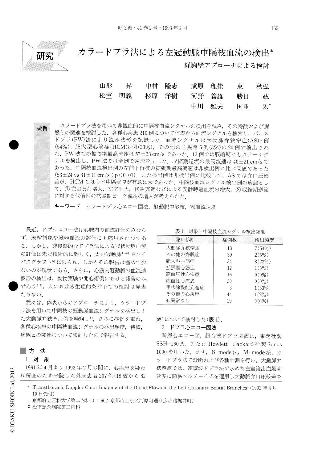
カラードプラ法を用いて非観血的に中隔枝血流シグナルの検出を試み,その特徴および病態との関連を検討した.各種心疾患210例について体表から血流シグナルを検索し,パルスドプラ(PW)法により流速波形を記録した.血流シグナルは大動脈弁狭窄症(AS)7例(54%),肥大型心筋症(HCM)8例(23%),その他の心異常5例(3%)の20例で検出された.PW法での拡張期最高流速は57±23cm/sであった.13例では収縮期にもカラーシグナルを検出し,PW法では全例で逆流を呈した.収縮期逆流の最高流速は40±21cm/sであった.中隔枝血流検出例の左前下行枝の拡張期最高流速は非検出例に比べ高値であった(53±24vs 31±11cm/s;p<0.01).また検出例は非検出例に比較して,ASでは弁口圧較差が,HCMでは心室中隔壁厚が有意に大であった.中隔枝血流シグナル検出例の病態として,①左室負荷増大,左室肥大,代謝亢進などによる安静時冠血流の増大,②収縮期逆流に対する代償性の拡張期ピーク流速の増大が考えられた.
This study characterized blood flow signals derived from the left coronary septal branches by transthoracic Doppler color flow imaging. In the anterior ventricular septum, the signal was detected in 7 of 13 patients with aortic stenosis, 8 of 34 with hypertrophic car-diomyopathy, and 5 of 144 patients with other diseased states. The peak diastolic flow velocity assessed by a pulsed Doppler technique ranged 21-115 cmis (mean 57). Systolic signal was depicted in 13 of the 20 with the diastolic signal, indicating retrograde flow direction in all of them. The peak negative systolic component ranged 11-80 cm/s (mean 40).

Copyright © 1993, Igaku-Shoin Ltd. All rights reserved.


