Japanese
English
- 有料閲覧
- Abstract 文献概要
- 1ページ目 Look Inside
- サイト内被引用 Cited by
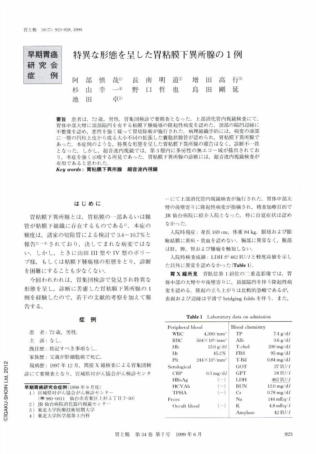
要旨 患者は,72歳,男性.胃集団検診で要精査となった.上部消化管内視鏡検査にて,胃体中部大彎に頂部陥凹を有する粘膜下腫瘍様の隆起性病変を認めた.頂部の陥凹辺縁に不整像を認め,悪性を強く疑って胃切除術が施行された.病理組織学的には,病変の深部に一層の円柱上皮から成る大小不同の拡張した囊胞状腺管が認められ,胃粘膜下異所腺であった.本症例のような,特異な形態を呈した胃粘膜下異所腺の報告はなく,診断不一致となった.しかし,超音波内視鏡では,第3層内に多房性の無エコー域が描出されており,本症を強く示唆する所見であった.胃粘膜下異所腺の診断には,超音波内視鏡検査が有用であると思われた.
A 72-year-old man was admitted to the JR Sendai hospital for further examination of his stomach, because he had been found, on the occasion of a gastric mass survey, to have an elevated lesion in his stomach. An upper gastrointestinal x-ray examination demonstrated a round elevated lesion with central depression in the middle gastric body. Endoscopic examination demonstrated a submucosal lesion with bridging folds and a pale colored depression was seen at the top of the lesion. Endoscopic ultrasonography demonstrated a multiple aechoic region with part of a hypoechoic region located in the third layer of the gastric wall. Histologically, the upper part of the lesion consisted of fibromuscular tissue and a relatively close gland. The deeper part of the lesion is consisted with the dilated glands like the pyloric gland. The endoscopic ultrasonographic findings reflected the histological findings well. Thus, it is concluded that ultrasonography is very useful for the diagnosis of submucosal heterotopia of gastric glands.
Copyright © 1999, Igaku-Shoin Ltd. All rights reserved.