Japanese
English
- 有料閲覧
- Abstract 文献概要
- 1ページ目 Look Inside
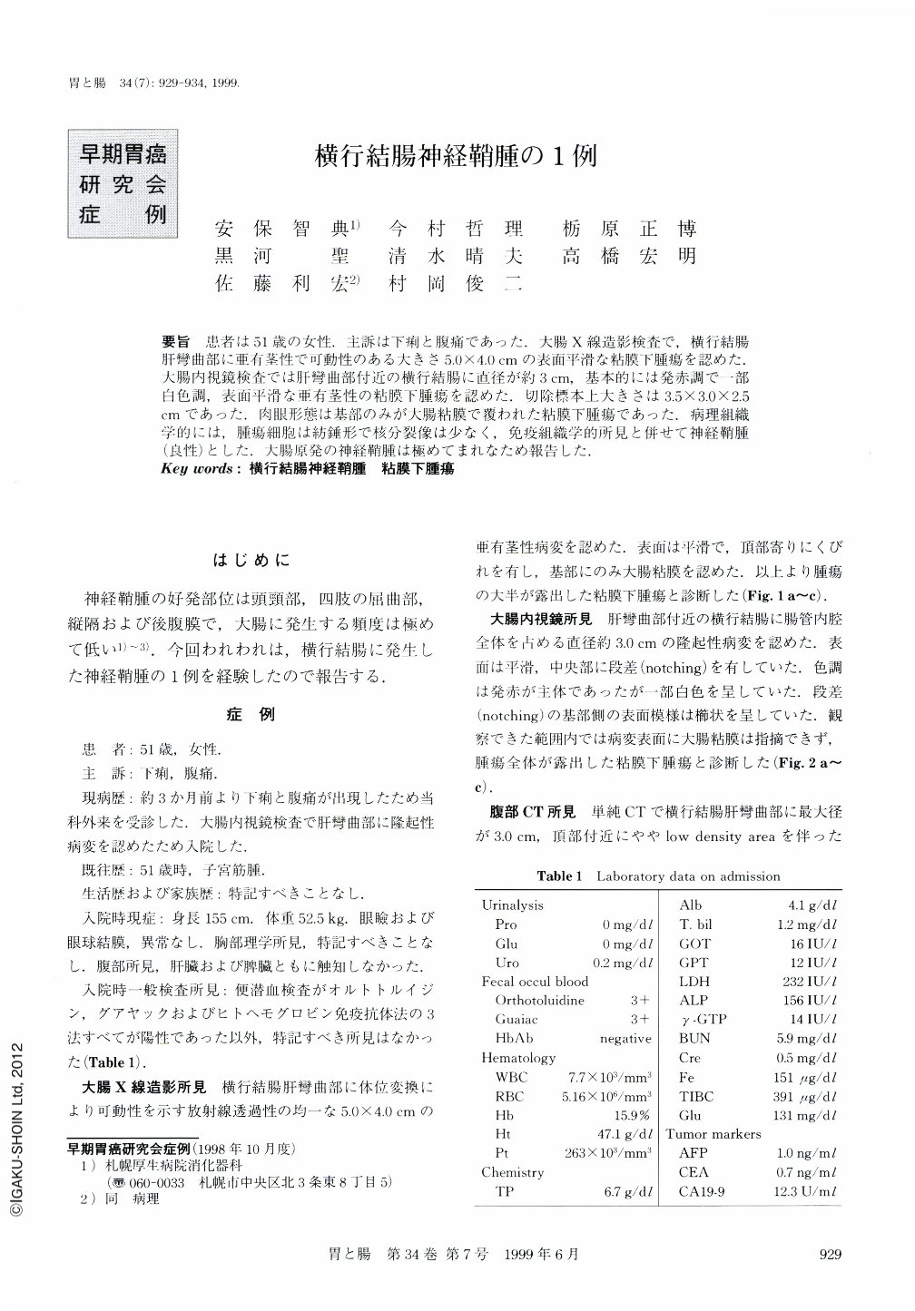
要旨 患者は51歳の女性.主訴は下痢と腹痛であった.大腸X線造影検査で,横行結腸肝彎曲部に亜有茎性で可動性のある大きさ5.0×4.0cmの表面平滑な粘膜下腫瘍を認めた.大腸内視鏡検査では肝彎曲部付近の横行結腸に直径が約3cm,基本的には発赤調で一部白色調,表面平滑な亜有茎性の粘膜下腫瘍を認めた.切除標本上大きさは35×3.0×2.5cmであった.肉眼形態は基部のみが大腸粘膜で覆われた粘膜下腫瘍であった.病理組織学的には,腫瘍細胞は紡錘形で核分裂像は少なく,免疫組織学的所見と併せて神経鞘腫(良性)とした.大腸原発の神経鞘腫は極めてまれなため報告した.
A neurilemoma is a tumor of the Schwann cell of the peripheral nervous system. It has a predilection for the head, neck and flexor surface of the upper and lower extremities, but a colonic one is extremely rare. So we reported a case of neurilemoma of the transverse colon as follows :
A 51-year-old female was admitted to our hospital because of diarrhea and abdominal pain. Double contrast colonography showed a homogeneous hyporadiolucent semipedunculated tumor in the hepatic flexure of the transverse colon, most of which, except for at the base, was not covered with colonic mucosa. Colonoscopic examination revealed a bulky red roundshaped tumor, which, as far as could be seen, was not covered with any colonic mucosa. These findings led to the clinical diagnosis of an exposed submucosal tumor in the tansverse colon. Partial colectomy was performed as a result of the clinical diagnosis. The size of the tumor on the resected specimen was 3.5 × 2.5 × 3.0 cm. Macroscopic view demonstrated a submucosal tumor though most of the surface of the tumor was not covered with colonic mucosa. Microscopic study confirmed that the tumor was composed of spindle-shaped cells with eosinophilic cytoplasm and ovoid nuculei arranged in pallisades. Immunohistochemical stain revealed that S-100 protein, glial fibrillary acidic protein (GFAP), Leu7, and vimentin were present, but smooth muscle actin, desmin, and CD34 were absent. The mitosis figures were scanty (17 per 4,500 high-power-fields) . Dysplasia was not detected clearly. Consequently, the diagnosis was a benign neurilemoma of the transverse colon.

Copyright © 1999, Igaku-Shoin Ltd. All rights reserved.


