Japanese
English
- 有料閲覧
PPV購入案内
1,320円 (1,200円+税10%) こちらは、821ページから825ページの文献です。購入には医書.jp本会員登録が必要です。
会員登録完了後に改めて上記「購入サイトへ」を選択してください。
または「購入サイトへ」選択後に「購入ログイン」、「新規会員登録」の順に進んでください。
会員登録について詳しくはをご確認ください。
- Abstract 文献概要
- 1ページ目 Look Inside
- 参考文献 Reference
- サイト内被引用 Cited by
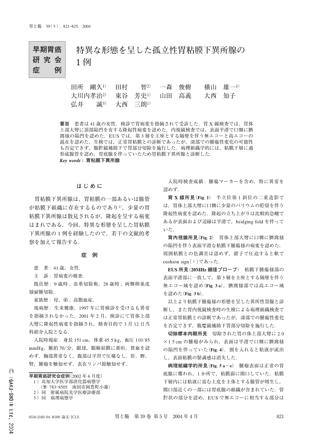
要旨 患者は41歳の女性.検診で胃病変を指摘されて受診した.胃X線検査では,胃体上部大彎に頂部陥凹を有する隆起性病変を認めた.内視鏡検査では,表面平滑で口側に臍窩様の陥凹を認めた.EUSでは,第3層を主座とする隔壁を伴う無エコーと高エコーの混在を認めた.生検では,正常胃粘膜との診断であったが,深部での腫瘍性変化の可能性も否定できず,腹腔鏡補助下で胃部分切除を施行した.病理組織学的には,粘膜下層に過形成腺管を認め,胃底腺を伴っていたため胃粘膜下異所腺と診断した.
A 41-year-old woman visited our hospital for further examination because she had been found to have an abnormality in her stomach during a medical check up. An upper gastrointestinal X-ray series demonstrated a round elevated lesion with bridging fold in the greater curvature of the upper gastric body. Endoscopic examination revealed a submucosal tumor-like lesion with smooth surface and Delle. Endoscopic ultrasonography demonstrated an anechoic region with septum and a hyperechoic part in the region located in the third layer of the gastric wall. In the biopsy, although it was diagnosed with normal stomach membrane, the possibility of neoplastic change in the deeper layer could not be denied, so partial gastrectomy was performed using laparoscopy. Histologically, its diagnosis was a submucosal heterotopia of the gastric gland, because hyperplastic glands with fundic glands were revealed in the submucosal layer.
1) Department of Gastroenterology and Hepatology, Kochi Medical School, Nankoku, Japan
2) Department of Endoscopy, Kochi Medical School, Nankoku, Japan
Copyright © 2004, Igaku-Shoin Ltd. All rights reserved.