Japanese
English
症例
MRI拡散強調像が感染性腎嚢胞の診断に有用であった1例
A case of infected renal cyst which was accurately diagnosed using MRI DWI:a case report
沼 哲也
1
,
山本 泰宏
1
,
松田 かおり
1
,
岡崎 良夫
1
,
木本 眞
1
,
平木 隆夫
2
Tetsuya Numa
1
1放射線第一病院 放射線科
2岡山大学病院 放射線科
1Department of Radiology Housyasen Daiichi Hospital
キーワード:
感染性腎嚢胞
,
拡散強調像
,
CTガイド下ドレナージ
Keyword:
感染性腎嚢胞
,
拡散強調像
,
CTガイド下ドレナージ
pp.821-825
発行日 2021年8月10日
Published Date 2021/8/10
DOI https://doi.org/10.18888/rp.0000001681
- 有料閲覧
PPV購入案内
1,650円 (1,500円+税10%) こちらは、821ページから825ページの文献です。
PPV(ペイ・パー・ビュー)とは、論文(記事)単位で購入・閲覧ができるサービスです。
購入には医書.jp本会員登録が必要です。
会員登録完了後に改めて上記「購入サイトへ」を選択してください。
または「購入サイトへ」選択後に「購入ログイン」、「新規会員登録」の順に進んでください。
会員登録について詳しくはをご確認ください。
購入には医書.jp本会員登録が必要です。
会員登録完了後に改めて上記「購入サイトへ」を選択してください。
または「購入サイトへ」選択後に「購入ログイン」、「新規会員登録」の順に進んでください。
会員登録について詳しくはをご確認ください。
- Abstract 文献概要
- 1ページ目 Look Inside
- 参考文献 Reference
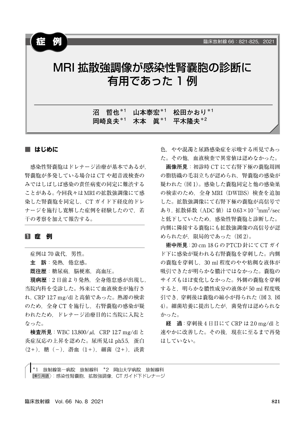
感染性腎嚢胞はドレナージ治療が基本であるが,腎嚢胞が多発している場合はCTや超音波検査のみではしばしば感染の責任病変の同定に難渋することがある。今回我々はMRIの拡散強調像にて感染した腎嚢胞を同定し,CTガイド下経皮的ドレナージを施行し寛解した症例を経験したので,若干の考察を加えて報告する。
We report a case of infected renal cyst with fever and sense of fatigue. CT showed right renal cysts surrounded by a hyperdense area of perirenal fat tissue. We suspected infected renal cysts and two of renal cysts showed high signal on MRI-DWI(Diffusion Weighted Imaging). We punctured the cysts and drained the fluid. One of them which showed partically high signal on MRI-DWI was serous drainage and the other which was high signal overall was purulent drainage. Two years later, the renal cyst did not recur.
Copyright © 2021, KANEHARA SHUPPAN Co.LTD. All rights reserved.