Japanese
English
原著
MRI拡散強調像が有用であった中耳真珠腫症例
The 2 cases of middle ear cholesteatoma diagnosed with diffusion-weighted MR imaging
菅原 一真
1
,
下郡 博明
1
,
橋本 誠
1
,
御厨 剛史
1
,
山下 裕司
1
Kazuma Sugahara
1
1山口大学大学院医学系研究科耳鼻咽喉科学分野
1Department of Otolaryngology,Yamaguchi University Graduate School of Medicine
pp.521-525
発行日 2009年7月20日
Published Date 2009/7/20
DOI https://doi.org/10.11477/mf.1411101459
- 有料閲覧
PPV購入案内
1,320円 (1,200円+税10%) こちらは、521ページから525ページの文献です。
PPV(ペイ・パー・ビュー)とは、論文(記事)単位で購入・閲覧ができるサービスです。
購入には医書.jp本会員登録が必要です。
会員登録完了後に改めて上記「購入サイトへ」を選択してください。
または「購入サイトへ」選択後に「購入ログイン」、「新規会員登録」の順に進んでください。
会員登録について詳しくはをご確認ください。
購入には医書.jp本会員登録が必要です。
会員登録完了後に改めて上記「購入サイトへ」を選択してください。
または「購入サイトへ」選択後に「購入ログイン」、「新規会員登録」の順に進んでください。
会員登録について詳しくはをご確認ください。
- Abstract 文献概要
- 1ページ目 Look Inside
- 参考文献 Reference
Ⅰ.はじめに
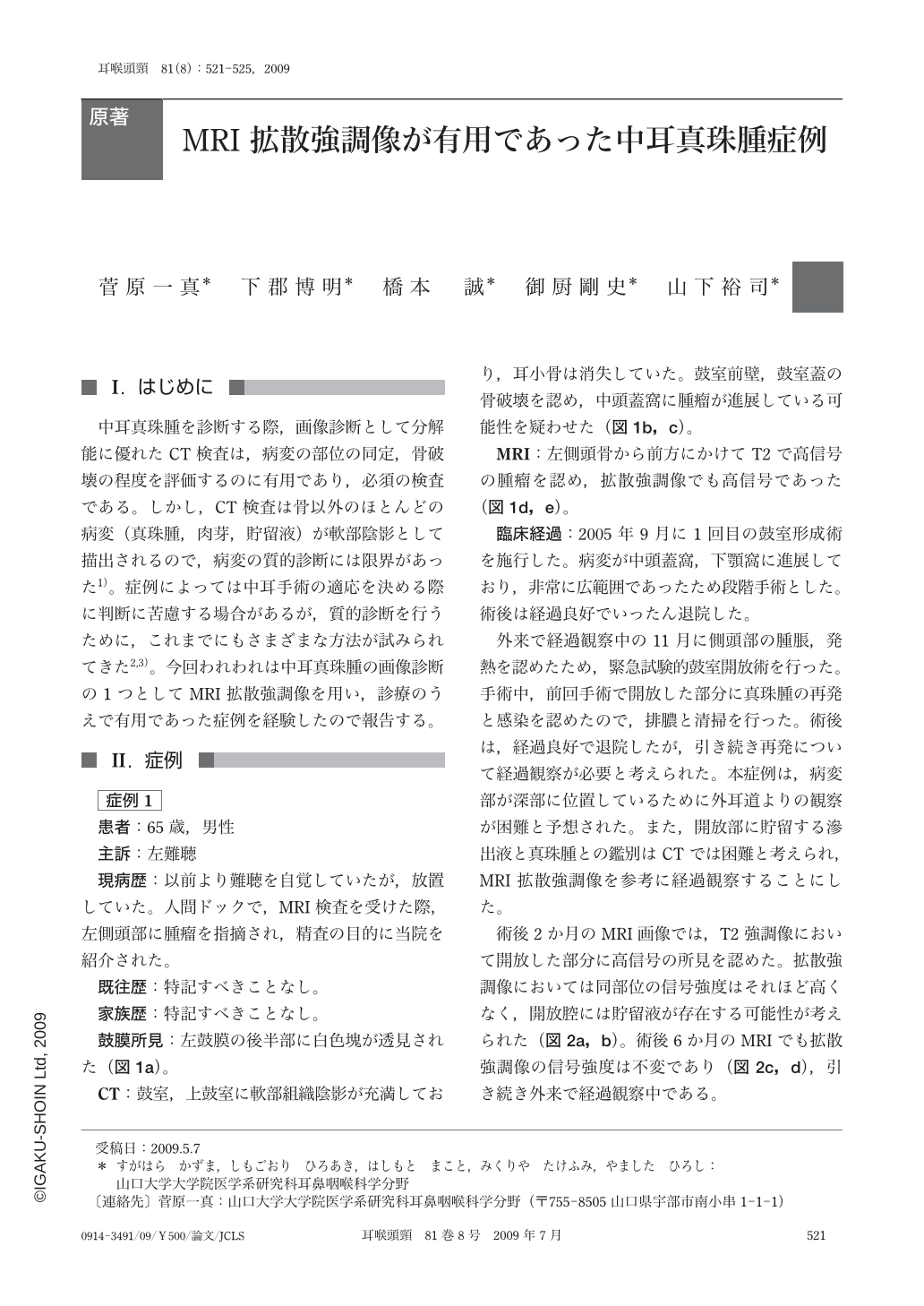
中耳真珠腫を診断する際,画像診断として分解能に優れたCT検査は,病変の部位の同定,骨破壊の程度を評価するのに有用であり,必須の検査である。しかし,CT検査は骨以外のほとんどの病変(真珠腫,肉芽,貯留液)が軟部陰影として描出されるので,病変の質的診断には限界があった1)。症例によっては中耳手術の適応を決める際に判断に苦慮する場合があるが,質的診断を行うために,これまでにもさまざまな方法が試みられてきた2,3)。今回われわれは中耳真珠腫の画像診断の1つとしてMRI拡散強調像を用い,診療のうえで有用であった症例を経験したので報告する。
We reported 2 patients who were diagnosed as middle ear cholesteatoma with diffusion-weighted MR imaging. In the first case,diffusion-weighted MR imaging was useful for checking the recurrence of cholesteatoma after surgery. In the second case,the imaging was helpful for the diagnosis of atypical case of cholesteatoma. This imaging method could be one of the important diagnostic techniques in otological field.
Copyright © 2009, Igaku-Shoin Ltd. All rights reserved.