Japanese
English
- 有料閲覧
PPV購入案内
1,320円 (1,200円+税10%) こちらは、1067ページから1069ページの文献です。購入には医書.jp本会員登録が必要です。
会員登録完了後に改めて上記「購入サイトへ」を選択してください。
または「購入サイトへ」選択後に「購入ログイン」、「新規会員登録」の順に進んでください。
会員登録について詳しくはをご確認ください。
- Abstract 文献概要
- 1ページ目 Look Inside
I.はじめに
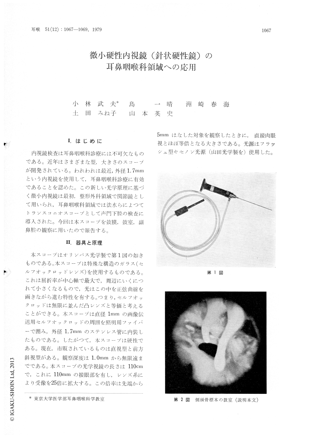
内視鏡検査は耳鼻咽喉科診療には不可欠なものである。近年はさまざまな型,大きさのスコープが開発されている。われわれは最近,外径1.7mmという内視鏡を使用して,耳鼻咽喉科診療に有効であることを認めた。この新しい光学原理に基づく微小内視鏡は最初,整形外科領域で関節鏡として用いられ,耳鼻咽喉科領域では法水らによつてトランスコニオスコープとして声門下腔の検査に導入された。今回は本スコープを鼓膜,鼓室,副鼻腔の観察に用いたので報告する。
A new slender type of endoscope is used for visualization of the tympanic membrane and inside of the maxillary sinus.
This scope is made of special rod lens (Selfoc).The Selfoc rod lens of 1.0 mm diameter is covered with glass fiber for illumination. The outer diameter of scope is only 1.7 mm. At present, two types of the scope are available : straight view type and anterior oblique type. This Selfoc scope is so slender that it can hardly be harmful.
Good candidates for ear scopy are those with the stenotic external canal. For inspection of the maxillary sinus, a trocar of 2.0 mm diameter is stabbed through the medial wall of the maxillary sinus at the inferior nasal meatus. Photography is easily taken using a powerful light source.
Copyright © 1979, Igaku-Shoin Ltd. All rights reserved.