Japanese
English
- 有料閲覧
PPV購入案内
1,320円 (1,200円+税10%) こちらは、641ページから644ページの文献です。購入には医書.jp本会員登録が必要です。
会員登録完了後に改めて上記「購入サイトへ」を選択してください。
または「購入サイトへ」選択後に「購入ログイン」、「新規会員登録」の順に進んでください。
会員登録について詳しくはをご確認ください。
- Abstract 文献概要
- 1ページ目 Look Inside
I.はじめに
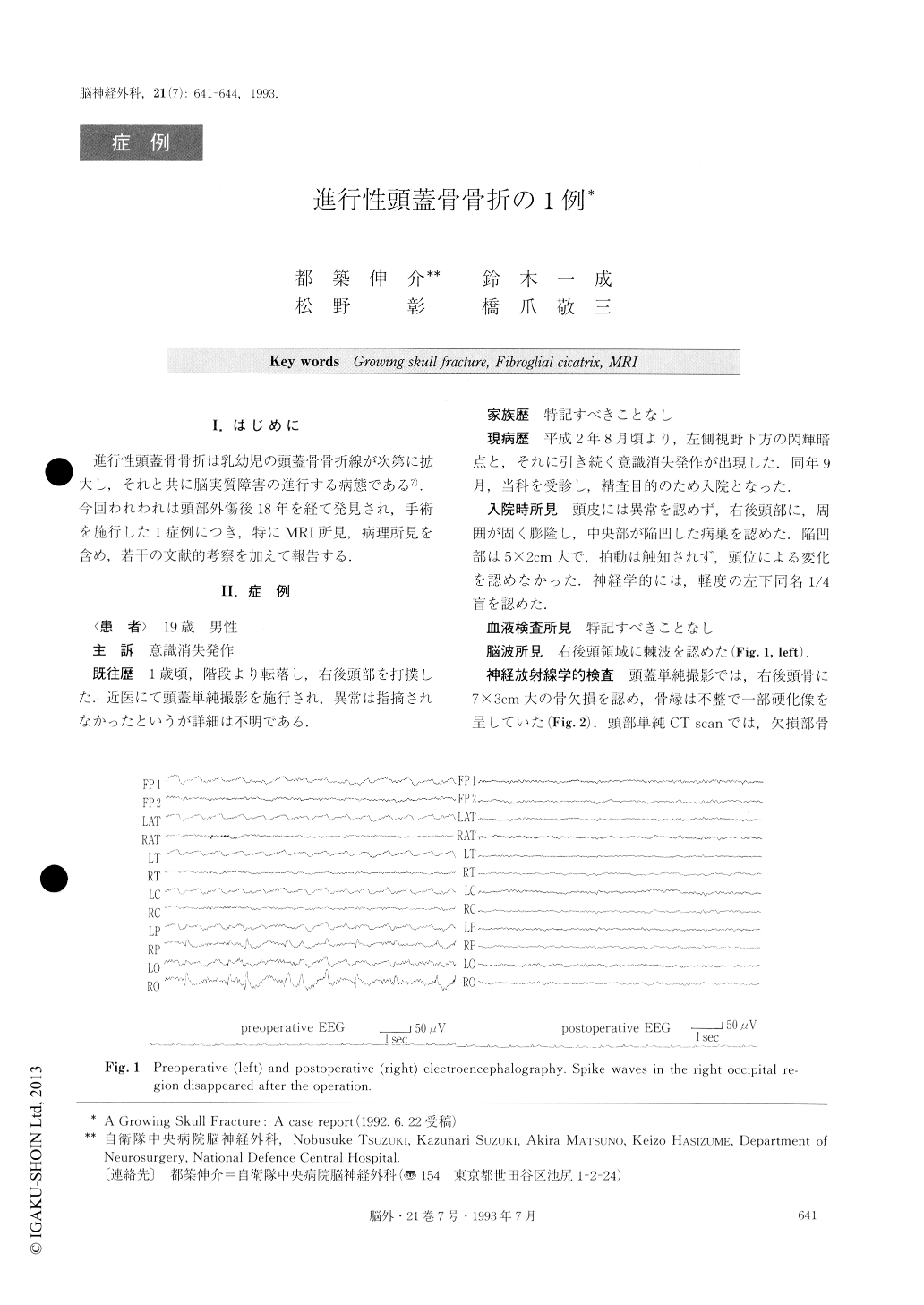
進行性頭蓋骨骨折は乳幼児の頭蓋骨骨折線が次第に拡大し,それと共に脳実質障害の進行する病態である7).今回われわれは頭部外傷後18年を経て発見され,手術を施行した1症例につき,特にMRI所見,病理所見を含め,若干の文献的考察を加えて報告する.
A case of growing skull fracture was reported with some considerations on the literature.
At the age of 1 year, the patient fell down stairs and struck the right occipital region, but (lid not receive any treatment at that time. At the age of 19 years, he suffered from several epileptic attacks and was admitted to our ward. Plain craniograms showed a right occipital bone defect measuring 7×3cm in size. CT scan demonstrated a cystic lesion just beneath a membranous substance ex-isting at the bone defect. MRI revealed cystic encephalo-malacia underlying the cyst very clearly. At the opera-tion the bone defect was found to be replaced by the gra-nulation tissue which was examined microscopically. Under the granulation tissue, a cyst filled with yellowish fluid and surrounding gliotic brain was found. The dural tear was noticed to be much wider than the bone defect. A dural plasty with LYODURAR and a cranioplasty with methacrylic resin were performed. Microscopic ex-amination revealed the granulation tissue to be a fibrog-lial cicatrix. According to these findings, we concluded that the contused and swollen brain tissue which had herniated through the dural tear had enlarged the skull fracture and formed a fibroglial cicatrix with sub-cutaneous tissue.
It is emphasized that MRI combined with CT scan is an indispensable procedure for diagnosis of growing skull fracture, and microscopic examination contributes to an understanding of its pathogenesis.
Copyright © 1993, Igaku-Shoin Ltd. All rights reserved.