Japanese
English
- 有料閲覧
PPV購入案内
1,320円 (1,200円+税10%) こちらは、1149ページから1151ページの文献です。購入には医書.jp本会員登録が必要です。
会員登録完了後に改めて上記「購入サイトへ」を選択してください。
または「購入サイトへ」選択後に「購入ログイン」、「新規会員登録」の順に進んでください。
会員登録について詳しくはをご確認ください。
- Abstract 文献概要
- 1ページ目 Look Inside
I.はじめに
成人慢性硬膜下血腫が両側に存在する場合,左右同時の穿頭血腫除去術が治療の原則である4).今回われわれは左右血腫腔が交通性を有し,一側のみの穿頭術で治癒した両側慢性硬膜下血腫の1例を経験したので,文献的考察を加えて報告する.
The authors present a case of bilateral chronic subdu-ral hematoma with communication between the hema-toma cavities.
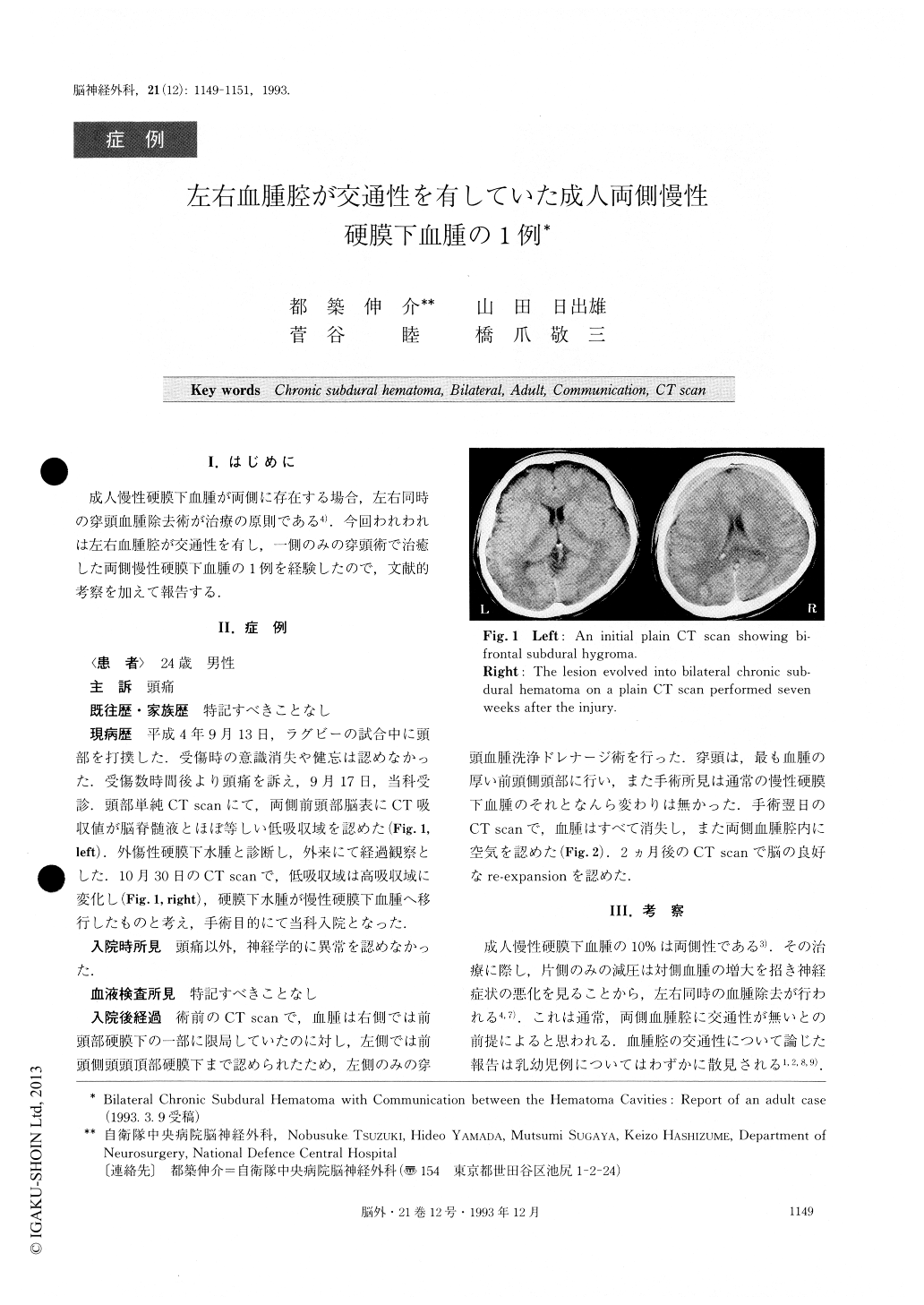
This 24-year-old male had hit his forehead while play-ing football and visited our hospital. An initial plain CT scan revealed extracerebral low density areas in the bifrontal region, which were considered to be post-traumatic subdural hygromas. The lesion was followed up with repeat CT scans. On plain CT scan performed seven weeks after the injury, the lesion had evolved into bilateral chronic subdural hematoma and the patient was admitted to our ward. He underwent burr hole drainage on the left side, bacause the hematoma volume on the left side was considerably larger than that on the right side. A plain CT scan on the day following the operation revealed complete resolution of the hematomas on both sides. A favorable re-expansion of the brain was con-firmed on consecutive CT scan performed two months after the operation.
In general bilateral chronic subdural hematomas in adults are considered to have no communication between the hematoma cavities and therefore they should be evacuated on both sides simultaneously. In our case, on the contrary, the operation revealed a communication be-tween the hematoma cavities.
The falx cerebri is narrow in front and this narrow anterior part is frequently perforated by one or more apertures. We attributed the communication to this anatom-ical feature of the falx cerebri. On preoperative CT scan, in addition, the inner surface of the bifrontal hema-toma cavity demonstrated a smooth concave figure in-dicating retrospectively that the cavity on the left side was continuous with that on the right side. Thus, meticu-lous investigation of CT scans is recommended in cases of bifrontal chronic subdural hematoma in adults, and when they demonstrate the figure mentioned above, treatment such as unilateral burr hole drainage may be applied.
Copyright © 1993, Igaku-Shoin Ltd. All rights reserved.