Japanese
English
- 有料閲覧
- Abstract 文献概要
- 1ページ目 Look Inside
I.はじめに
Growing skull fractureは乳幼児の頭蓋骨骨折が進行性に拡大する比較的まれな疾患である.その機序は骨折部直下のくも膜嚢胞14)あるいは脱出脳7,13,15)が頭蓋内の圧の拍動を骨折線に伝達し骨折線が拡大していくとされている.その経過は様々で数時間で骨折線の拡大を示す例4)から10年以上かけて進行する拡大骨折の例16)も報告されている.今回われわれは18生日の乳児で頭部外傷後,急速に骨折線の拡大を来たした例を経験した.その特徴的CT,MRI像を示し,growing skull fractureの治療について考察した.
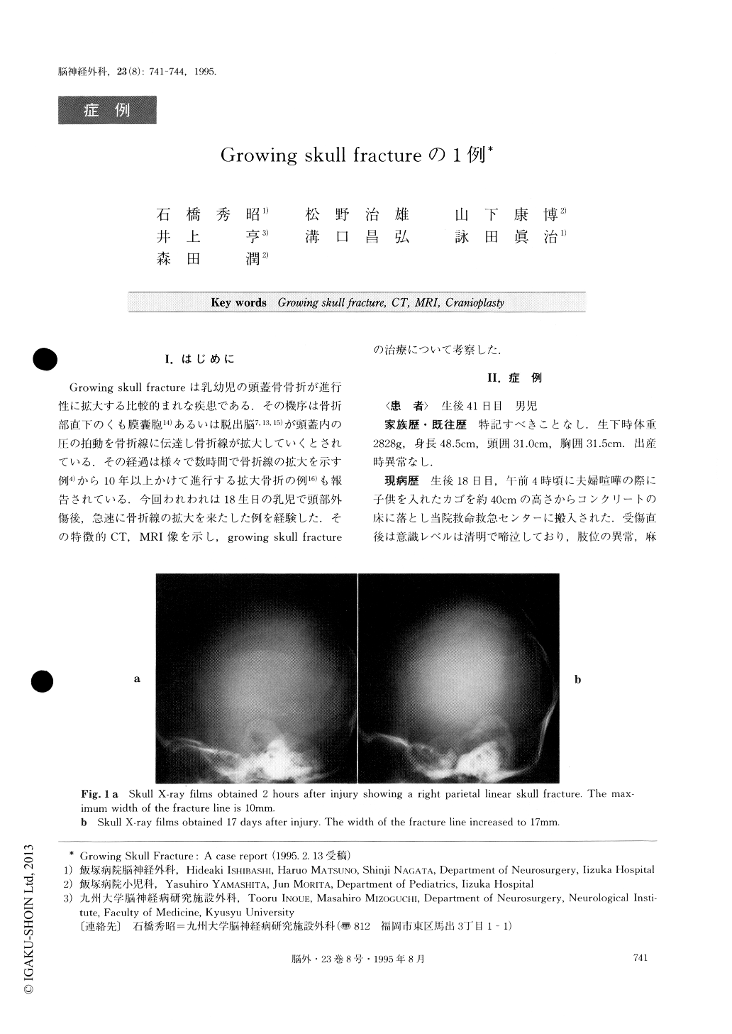
An 18-day-old male baby who had fallen from his mothers arms and hit his head on the floor was admit-ted to our hospital. On admission, the patient was crying, but no weakness was noted in the extremities. A small fluctuant protrusion was visible in the right parietal region. The plain skull X-ray film revealed a wide linear fracture in the parietal bone. Computed tomography (CT) showed swelling of the right hemi-sphere and a traumatic subarachnoid hemorrhage. At 41 days old, the subcutaneous fluid collection had in-creased in volume and the width of the linear skull fracture was also enlarged as shown on the X-ray film.CT and magnetic resonance imaging (MRI) revealed a large cyst herniating through the wide parietal bone de-fect. There was also an enlarged right lateral ventricle and a torn clural margin in the brain. The cranioplasty with dural plasty was performed on the 43th clay of ago under the diagnosis of growing skull fracture of the right parietal bone. The postoperative course was uneventful, without seizure or weakness.
In order to diagnose growing skull fracture, especial-ly to show the relationship between the fracture, torn dura matter, the ventricle and the contused brain, MRI was very helpful combined with CT and plain skull X-rays. Cranioplasty with clural repair was considered the essential procedure for the treatment of such growing skull fractures.

Copyright © 1995, Igaku-Shoin Ltd. All rights reserved.


