Japanese
English
- 有料閲覧
PPV購入案内
1,320円 (1,200円+税10%) こちらは、935ページから939ページの文献です。購入には医書.jp本会員登録が必要です。
会員登録完了後に改めて上記「購入サイトへ」を選択してください。
または「購入サイトへ」選択後に「購入ログイン」、「新規会員登録」の順に進んでください。
会員登録について詳しくはをご確認ください。
- Abstract 文献概要
- 1ページ目 Look Inside
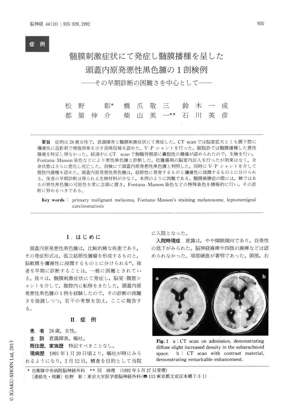
症例は28歳女性で,意識障害と髄膜刺激症状にて発症した。CT scanでは脳室拡大とくも膜下腔に瀰漫性に造影剤で増強効果を示す高吸収域を認めた。V-Pシャントを行った。細胞診では髄膜播種した悪性腫瘍を特定し得なかった。経過中にCT scanで胸髄背側部に嚢胞性の腫瘍が認められたので,生検を行い,Fontana Masson染色などにより悪性黒色腫と診断した。抗腫瘍剤の脳室内注入を行ったが効果はなく,全身状態はさらに悪化し死亡した。剖検にて頭蓋内原発悪性黒色腫と判明した。同時にV-Pシャントを介して腹腔内播種を認めた。頭蓋内原発悪性黒色腫は,結節性に発育するものと瀰漫性に浸潤するものとに分けられる。後者の早期診断は得られる生検材料が少なく,本例のように困難である。髄膜癌腫症の際には,稀ではあるが悪性黒色腫の可能性を常に念頭に置き,Fontana-Masson染色などの特殊染色を積極的に行い,その診断に努めるべきである。
A 28-year-old woman was hospitalized in drowsy state with signs of increased intracranial pressure. CT scans revealed diffuse increased density with marked enhancement in the subarachnoid space, as well as ventricular dilatation. V-P shunt operation was performed to control intracranial pressure. Repeated cytological examinations of CSF couldn't determine the tumor origin. CT scan of thoracic spine showed a cystic tumor in its dorsal aspect. T2-weighted MRI revealed multiple spotty low inten-sity, specific to melanin granules, throughout the whole spine. Her thoracic spine was explored, and the intradural tumor was partially removed. His-topathological examination revealed the tumor cell which had dark nucleus with conspicuous nucleolus and cytoplasmic granules. These findings were compatible with malignant melanoma. Her general condition were deteriorated progressively and she died about 5 months after her admission. Postmor-turn examination showed diffuse leptomeningeal invasion of dark tumor throughout the entire cen-tral nervous system, and metastasis to peritoneum and omentum via V-P shunt system. Histopath-ological examination proved the tumor to be malig-nant melanoma. Electrone microscopic examina-tion also revealed melanosome in the cytoplasm.
Primary intracranial malignant melanoma is devided in two groups, nodular type and leptomenin-geal type. In the latter type, early diagnosis is very difficult, just as in our case, because only a little tissue specimen can be obtained. In a case of le-ptomenigeal carcinomatosis, possibility of primary malignant melanoma, though rare, should always be kept in mind, and specific staining such as Fontana -Masson's staining should be tried.
Copyright © 1992, Igaku-Shoin Ltd. All rights reserved.