Japanese
English
- 有料閲覧
- Abstract 文献概要
- 1ページ目 Look Inside
I.はじめに
Gliosarcomaはanaplastic astrocytomaあるいは神経膠芽腫の2-8%の頻度に併在する4,7,8),頭痛,視力障害及びうっ血乳頭で発症し,大きな嚢胞を伴い壁在結節様を有するgliosarcomaの1例を経験したので臨床像,組織学的診断および画像診断について,文献的考察を加え報告する.
A case of gliosarcoma with a large cyst is reported. A 22-year-old female was admitted to our hospital with complaints of blurred vision and headache.
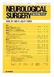
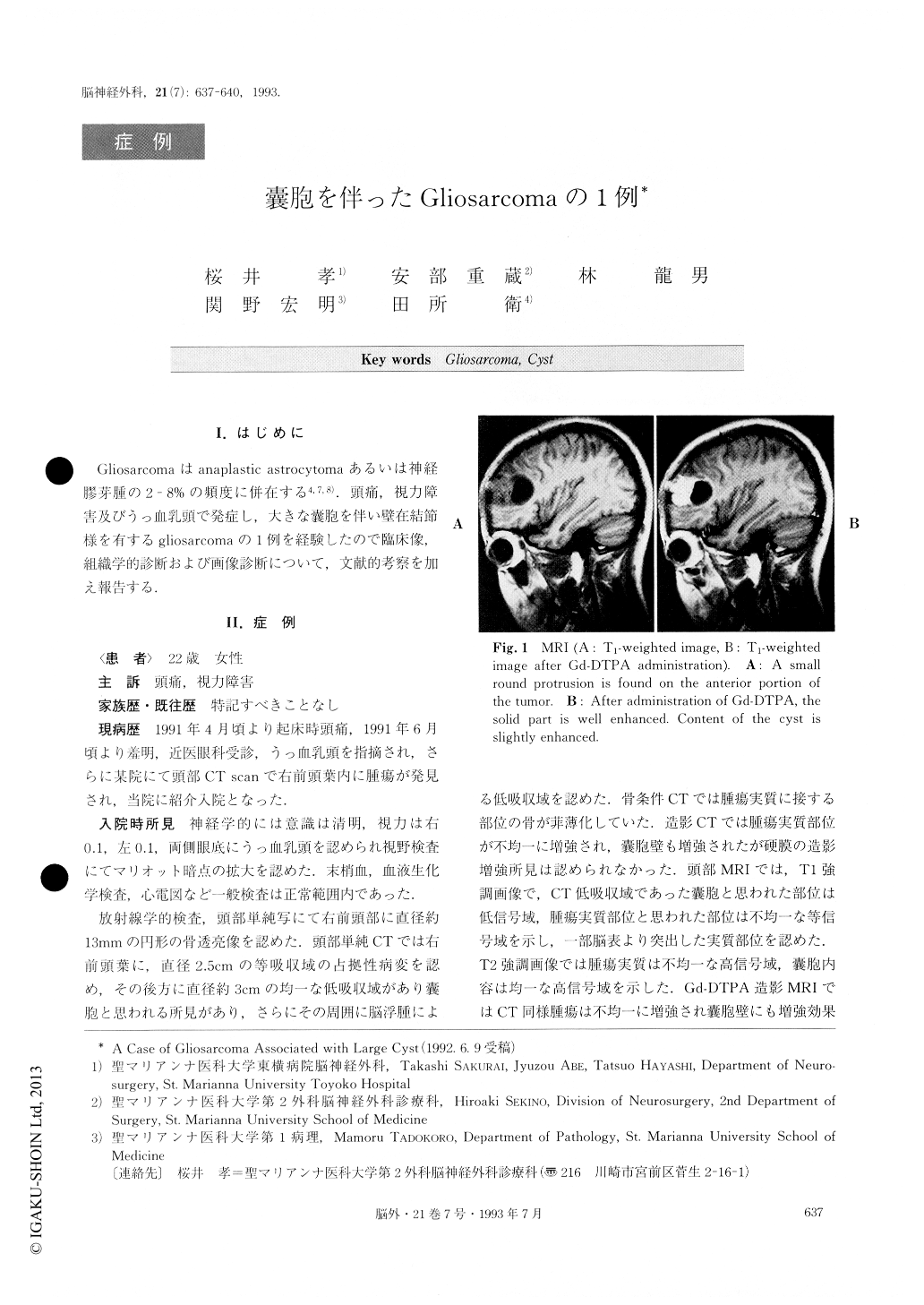
Plain skull x-ray films showed a radiolucent area in the right frontal area. Computed tomography (CT) revealed an iso-dense mass in the right frontal lobe with a large cyst. After administration of contrast medium, the solid part and cyst wall were well enhanced and the content of the cyst was slightly enhanced. CT number of the cyst fluid was increased from 64.2 to 83.5 Hounsfield units, after administration of the contrast medium. Axial T1-weighted magnetic resonance image (MRI) revealed an iso-intense mass with marked enhancement by Gd-DTPA in the same area. A large cyst was shown to be located in the dorsal part of the mass. A small round protrusion, 10mm in diameter, was found on the anterior portion of the mass on this MRI. Right carotid angiogram showed a tumor stain fed by the frontopolar artery. Right frontal lobectomy including the tumor was carried out with a preoperative diagnosis of glioblastoma. The patient received radiation therapy of 6OGy (whole brain 40Gy ; focal 20Gy) and chemotherapy postoperatively.
Histologically, necrosis, hemorrhage and endothelial hyperplasia were revealed at the tumor lesion. The tumor was composed of proliferation of glial and mesenchymal elements. The glial element appeared as fibrillary astrocytoma and polar spongioblastoma. The mesenchymal element showed sarcoma. As mentioned above, this tumor was diagnosed as gliosarcoma. It was difficult to make a diagnosis of gliosarcotna preoperatively because of the complex findings similar to malignant gliomas in conventional neuroradiological imaging. Generally, it is reported that the gliosarcoma is estimated to occur in from 2 to 8% of malignant gliomas. However, a gliosarcoma associated with a large cyst is quite rare. Considering radiological and histological findings, the contrast medium appeared to pass through the vascular channels surrounding the cyst wall. Consequently, we speculate that extravasation is the mechanism leading to the formation of this large cyst.
Copyright © 1993, Igaku-Shoin Ltd. All rights reserved.