Japanese
English
- 有料閲覧
PPV購入案内
1,320円 (1,200円+税10%) こちらは、773ページから777ページの文献です。購入には医書.jp本会員登録が必要です。
会員登録完了後に改めて上記「購入サイトへ」を選択してください。
または「購入サイトへ」選択後に「購入ログイン」、「新規会員登録」の順に進んでください。
会員登録について詳しくはをご確認ください。
- Abstract 文献概要
- 1ページ目 Look Inside
I.はじめに
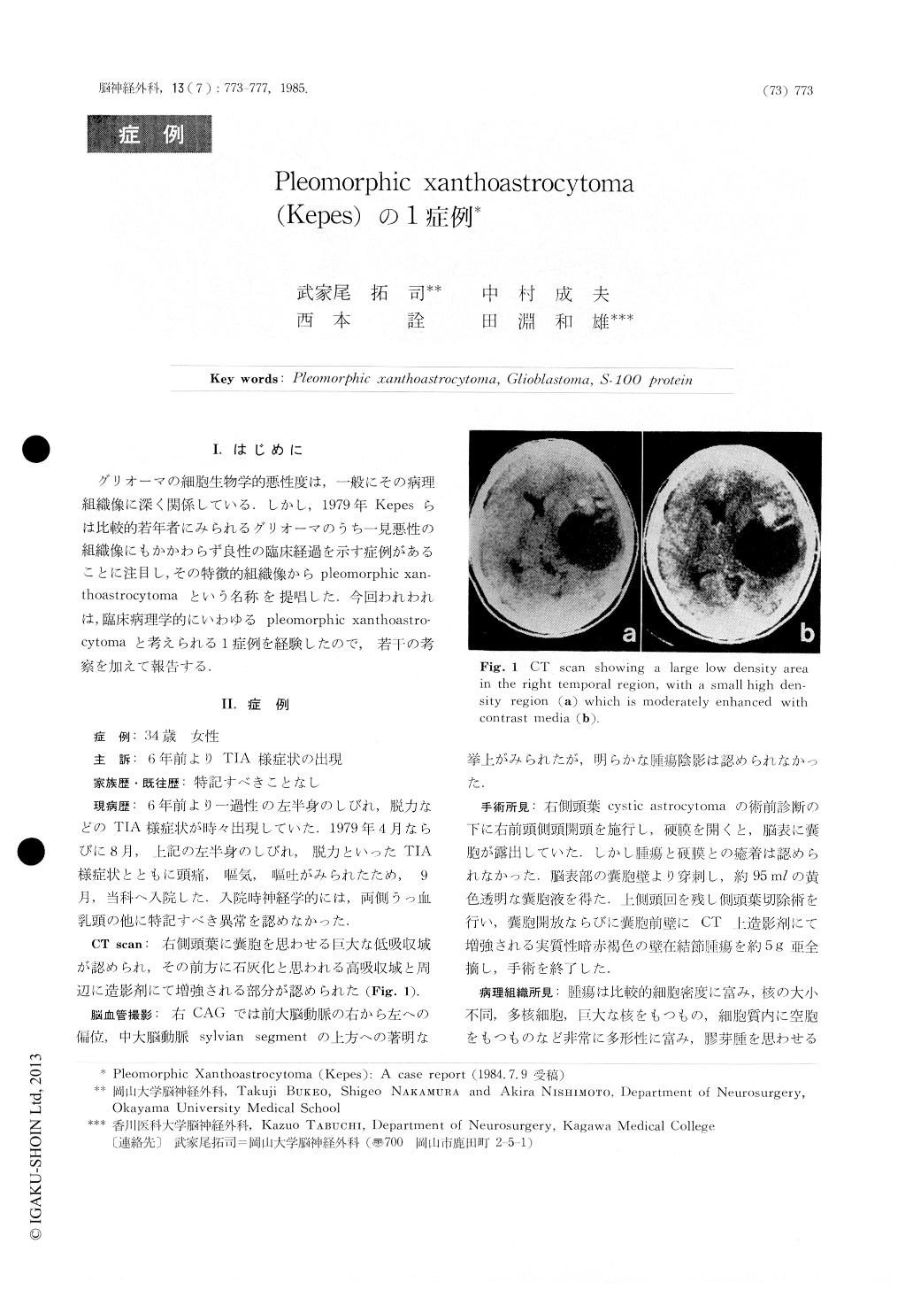
グリオーマの細胞生物学的悪性度は,一般にその病理組織像に深く関係している.しかし,1979年Kepesらは比較的若年者にみられるグリオーマのうち一見悪性の組織像にもかかわらず良性の臨床経過を示す症例があることに注目し,その特徴的組織像からpleomorphic xanthoastrocytomaという名称を提唱した.今回われわれは,臨床病理学的にいわゆるpleomorphic xanthoastrocytomaと考えられる1症例を経験したので,若干の考察を加えて報告する.
A case of tumor to be diagnosed as pleomorphic xanthoastrocytoma (Kepes) is reported. This patient was a 34-year-old female with a 6-year history of TIA. Neurological examination on admission showed no abnormalities except for bilateral choked disc. Plain CT scan revealed a well-defined low density area with asmall high dlensity region in the right temporal lobe. The small high density region and a part of peripheral portion of low density area were moderately enhanced with contrast media. At operation there was a cyst containing xanthochromic fluid at 1.5 cm depth from the cerebral surface.
Copyright © 1985, Igaku-Shoin Ltd. All rights reserved.