Japanese
English
- 有料閲覧
PPV購入案内
1,320円 (1,200円+税10%) こちらは、586ページから590ページの文献です。購入には医書.jp本会員登録が必要です。
会員登録完了後に改めて上記「購入サイトへ」を選択してください。
または「購入サイトへ」選択後に「購入ログイン」、「新規会員登録」の順に進んでください。
会員登録について詳しくはをご確認ください。
- Abstract 文献概要
- 1ページ目 Look Inside
- 参考文献 Reference
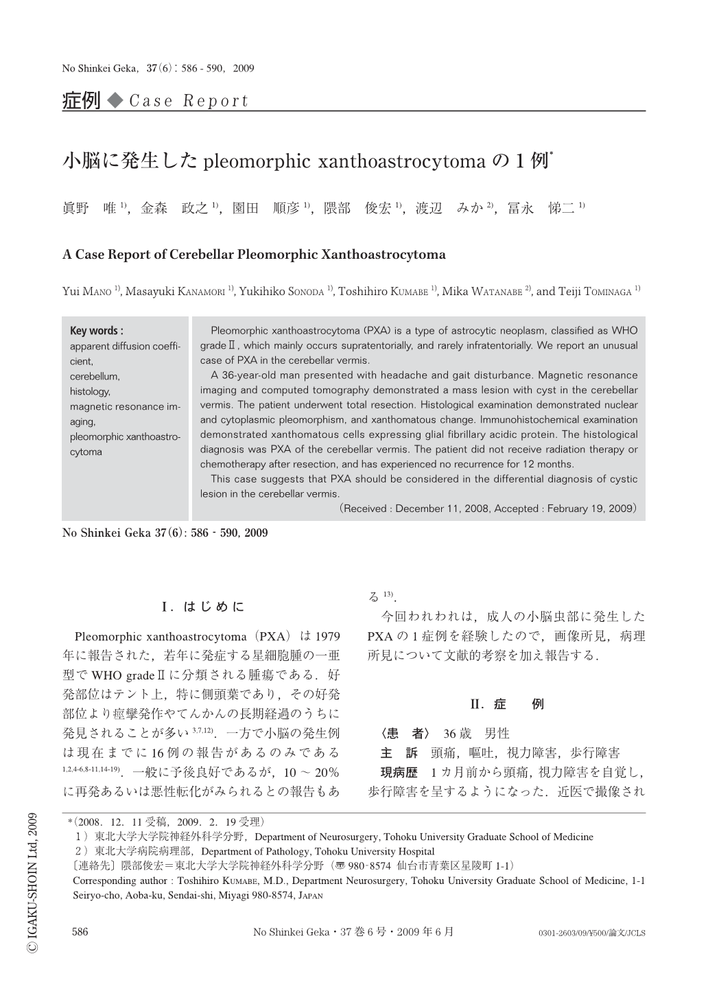
Ⅰ.はじめに
Pleomorphic xanthoastrocytoma(PXA)は1979年に報告された,若年に発症する星細胞腫の一亜型でWHO gradeⅡに分類される腫瘍である.好発部位はテント上,特に側頭葉であり,その好発部位より痙攣発作やてんかんの長期経過のうちに発見されることが多い3,7,12).一方で小脳の発生例は現在までに16例の報告があるのみである1,2,4-6,8-11,14-19).一般に予後良好であるが,10~20%に再発あるいは悪性転化がみられるとの報告もある13).
今回われわれは,成人の小脳虫部に発生したPXAの1症例を経験したので,画像所見,病理所見について文献的考察を加え報告する.
Pleomorphic xanthoastrocytoma (PXA) is a type of astrocytic neoplasm, classified as WHO gradeⅡ, which mainly occurs supratentorially, and rarely infratentorially. We report an unusual case of PXA in the cerebellar vermis.
A 36-year-old man presented with headache and gait disturbance. Magnetic resonance imaging and computed tomography demonstrated a mass lesion with cyst in the cerebellar vermis. The patient underwent total resection. Histological examination demonstrated nuclear and cytoplasmic pleomorphism, and xanthomatous change. Immunohistochemical examination demonstrated xanthomatous cells expressing glial fibrillary acidic protein. The histological diagnosis was PXA of the cerebellar vermis. The patient did not receive radiation therapy or chemotherapy after resection, and has experienced no recurrence for 12 months.
This case suggests that PXA should be considered in the differential diagnosis of cystic lesion in the cerebellar vermis.
Copyright © 2009, Igaku-Shoin Ltd. All rights reserved.